Suhandi Cecep, Wilar Gofarana, Mohammed Ahmed Fouad Abdelwahab, Mahmoud Safwat A, Muchtaridi Muchtaridi, Shamsuddin Shaharum, Safuan Sabreena, Lesmana Ronny, Hasan Nurhasni, Zulhendri Felix, Wathoni Nasrul
Doctoral Program of Pharmacy, Faculty of Pharmacy, Universitas Padjadjaran, Sumedang, West Java, 45363, Indonesia.
Department of Pharmaceutics and Pharmaceutical Technology, Faculty of Pharmacy, Universitas Padjadjaran, Sumedang, West Java, 45363, Indonesia.
J Inflamm Res. 2025 Jun 9;18:7443-7457. doi: 10.2147/JIR.S525243. eCollection 2025.
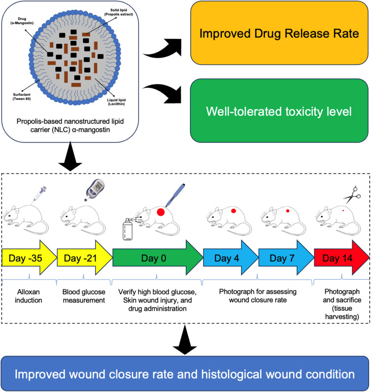
Diabetic wounds present a significant challenge due to delayed healing and susceptibility to infection. Conventional therapies often fall short of achieving complete and timely wound repair. This study investigates the potential of α-mangostin (αM) and its propolis-based nanostructured lipid carrier (NLC-P-αM) formulation as novel therapeutic agents for diabetic wound healing.
To evaluate the release profile, safety, and efficacy of NLC-P-αM in promoting wound repair in an in vitro and in vivo diabetic wound model.
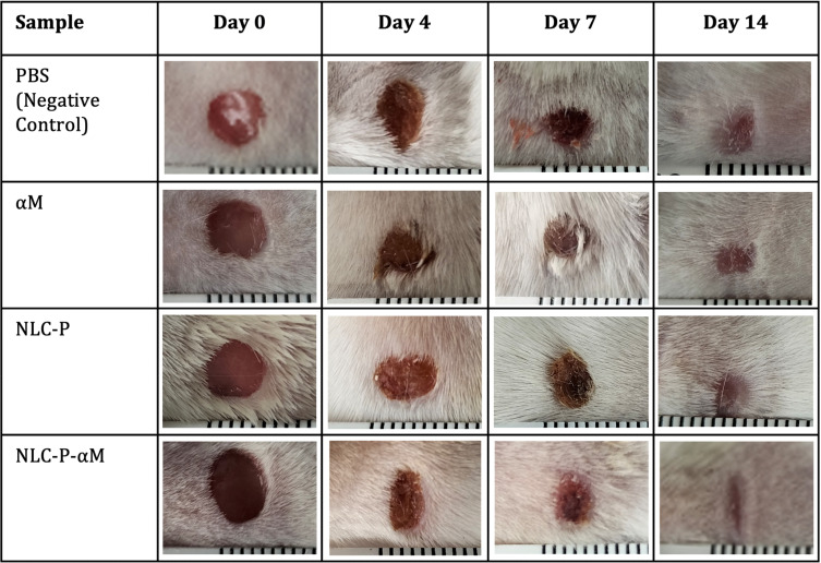
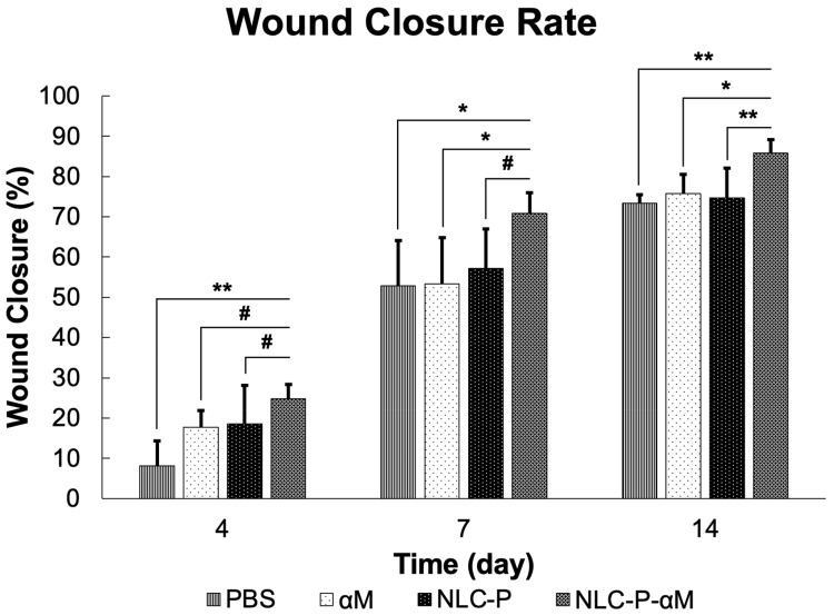
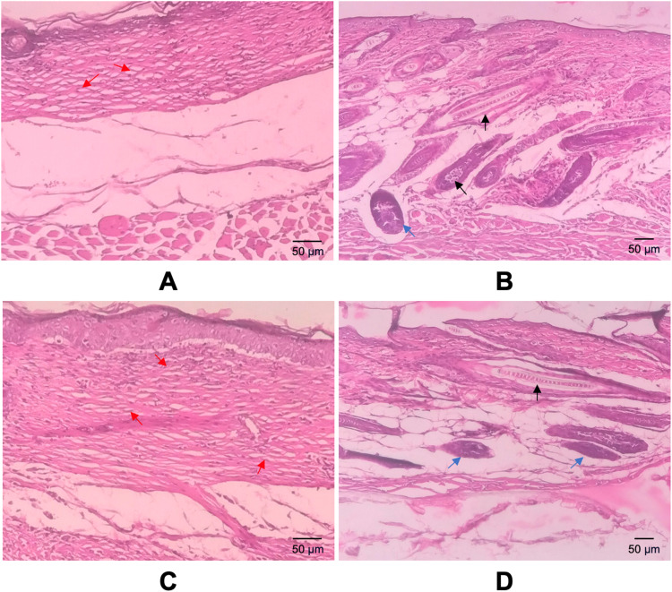
The NLC-P-αM formulation was prepared using a melt-emulsification technique with ultrasonication. In vitro release studies were conducted using a dialysis bag method and analyzed using kinetic models. Cytotoxicity was assessed using the WST-8 assay on NIH-3T3 fibroblast cells. In vivo diabetic wound healing was evaluated using alloxan-induced diabetic Swiss Webster mice. The treatments were applied topically for 14 days, and wound closure was monitored quantitatively. Histological analysis was performed to assess the inflammatory cell infiltration, epidermal thickness, and tissue regeneration.
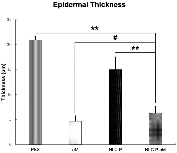
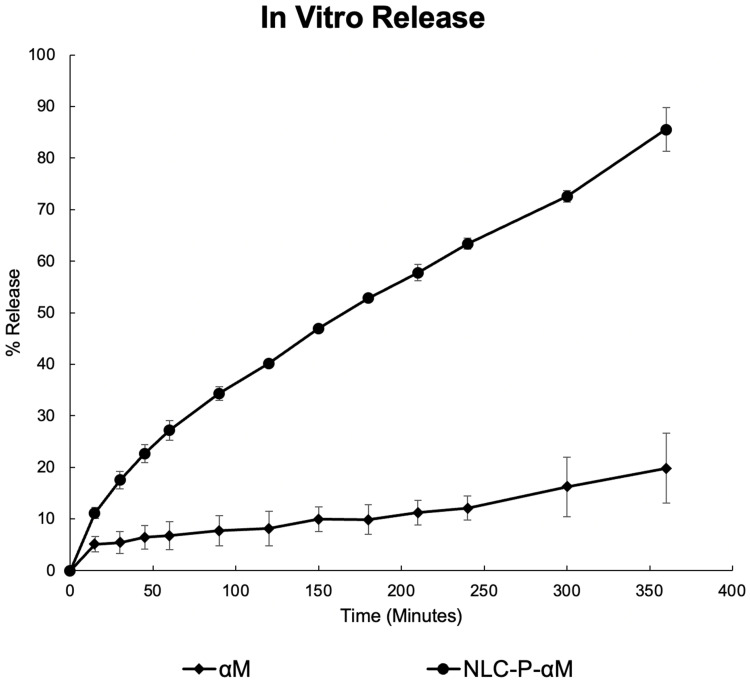
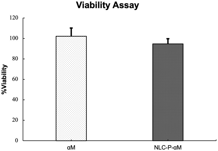
NLC-P-αM demonstrated a significantly enhanced release profile, with 85.55 ± 4.25% of αM released at 360 min compared to 19.82 ± 6.78% for free αM, following a non-Fickian diffusion mechanism. Both formulations exhibited excellent safety, with cell viabilities of 94.76 ± 4.95% for NLC-P-αM and 102.16 ± 7.98% for αM in NIH-3T3 cells. In vivo, NLC-P-αM achieved the highest wound closure rate (85.83 ± 3.33%) by day 14, outperforming αM and the controls. Histological analysis confirmed reduced inflammation, a thinner epidermis, and advanced tissue regeneration in the NLC-P-αM group, highlighting its superior therapeutic efficacy.
NLC-P-αM demonstrated enhanced release, excellent safety, and superior efficacy in promoting diabetic wound healing compared to free αM and other controls. This nanoformulation offers a promising therapeutic strategy for accelerating wound repair in diabetic patients.
糖尿病伤口由于愈合延迟和易感染而带来重大挑战。传统疗法往往难以实现完全及时的伤口修复。本研究调查了α-山竹素(αM)及其基于蜂胶的纳米结构脂质载体(NLC-P-αM)制剂作为糖尿病伤口愈合新型治疗剂的潜力。
评估NLC-P-αM在体外和体内糖尿病伤口模型中促进伤口修复的释放特性、安全性和疗效。
采用熔融乳化技术结合超声处理制备NLC-P-αM制剂。使用透析袋法进行体外释放研究,并采用动力学模型进行分析。使用WST-8法评估对NIH-3T3成纤维细胞的细胞毒性。使用四氧嘧啶诱导的糖尿病瑞士韦伯斯特小鼠评估体内糖尿病伤口愈合情况。局部应用治疗14天,定量监测伤口闭合情况。进行组织学分析以评估炎症细胞浸润、表皮厚度和组织再生情况。
NLC-P-αM表现出显著增强的释放特性,在360分钟时αM的释放量为85.55±4.25%,而游离αM为19.82±6.78%,遵循非菲克扩散机制。两种制剂均表现出优异的安全性,NLC-P-αM在NIH-3T3细胞中的细胞活力为94.76±4.95%,αM为102.16±7.98%。在体内,到第14天NLC-P-αM实现了最高的伤口闭合率(85.83±3.33%),优于αM和对照组。组织学分析证实NLC-P-αM组炎症减轻、表皮变薄且组织再生进展良好,突出了其卓越的治疗效果。
与游离αM和其他对照组相比,NLC-P-αM在促进糖尿病伤口愈合方面表现出增强的释放、优异的安全性和卓越的疗效。这种纳米制剂为加速糖尿病患者伤口修复提供了一种有前景的治疗策略。