Eagan Tomas M, Nielsen Rune, Haaland Ingvild, Husebø Gunnar R, Lehmann Sverre, Ward Jon A, Wilson Susan J
Department of Thoracic Medicine, Haukeland University Hospital, Bergen, Norway.
Department of Clinical Science, Faculty of Medicine, University of Bergen, Bergen, Norway.
PLoS One. 2025 Jun 17;20(6):e0326267. doi: 10.1371/journal.pone.0326267. eCollection 2025.
The understanding of inflammation and remodeling in the bronchial wall of COPD patients with varying disease severity remains incomplete.
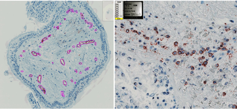
35 healthy controls and 47 volunteer COPD patients underwent bronchoscopy with bronchoalveolar lavage (BAL) and sampling by endobronchial biopsies in 2014-2015 as part of the MicroCOPD Study. Biopsies were embedded in glycol methyl acrylate (GMA) resin and examined by immunohistochemistry and staining for enumeration of CD3 + , CD4 + , CD8 + , CD20 + , CD68 + , EG2 + , and NE+ inflammatory cells, as well as endothelial cells (EN4) and smooth muscle actin (SMA). Mucus cells were stained by periodic acid-schiff (PAS), and toluidine blue to visualize the reticular basement membrane (RBM).
The numbers of macrophages and eosinophils were higher, and vascularity increased in the submucosa in COPD patients compared with healthy controls. In healthy smokers there were lower numbers of lymphocytes (CD3 +, CD4 +, CD8 +, CD20+) than never smokers. However, COPD patients with GOLD I/II had higher numbers of eosinophils and larger smooth muscle area compared with GOLD III/IV. COPD exacerbations the last year, blood eosinophils, and use of inhaled corticosteroids did not affect levels of inflammation or remodeling.
Smoking alters inflammation in healthy controls, while specific patterns of macrophages, eosinophils, and vascularity distinguish COPD from non-COPD in bronchial biopsies.
对于不同疾病严重程度的慢性阻塞性肺疾病(COPD)患者支气管壁炎症和重塑的理解仍不完整。
作为MicroCOPD研究的一部分,35名健康对照者和47名COPD志愿者在2014年至2015年接受了支气管镜检查,包括支气管肺泡灌洗(BAL)和经支气管活检取样。活检组织包埋于甲基丙烯酸乙二醇酯(GMA)树脂中,通过免疫组织化学和染色来计数CD3 +、CD4 +、CD8 +、CD20 +、CD68 +、EG2 +和NE +炎症细胞,以及内皮细胞(EN4)和平滑肌肌动蛋白(SMA)。黏液细胞用高碘酸 - 希夫(PAS)染色,甲苯胺蓝染色以观察网状基底膜(RBM)。
与健康对照者相比,COPD患者巨噬细胞和嗜酸性粒细胞数量增加,黏膜下层血管增多。健康吸烟者的淋巴细胞(CD3 +、CD4 +、CD8 +、CD20 +)数量低于从不吸烟者。然而,与GOLD III/IV级COPD患者相比,GOLD I/II级患者嗜酸性粒细胞数量更多,平滑肌面积更大。过去一年的COPD急性加重、血液嗜酸性粒细胞水平及吸入性糖皮质激素的使用并未影响炎症或重塑水平。
吸烟改变健康对照者的炎症状态,而巨噬细胞、嗜酸性粒细胞和血管的特定模式在支气管活检中可区分COPD与非COPD。