Liu Chengdong, Ding Yun, Yang Yiyuan, Huang Kenan, Wei Rongqiang, Xu Zhifei, Cai Zhigang, Tang Hua
Department of Thoracic Surgery, Shanghai Changzheng Hospital Affiliated to Second Military Medical University, Shanghai, China.
Department of Thoracic Surgery, Naval Medical Center of PLA, Shanghai, China.
J Thorac Dis. 2025 May 30;17(5):3307-3317. doi: 10.21037/jtd-2025-623. Epub 2025 May 15.
The management of esophageal anastomotic fistula continues to be a significant clinical challenge in the field of esophageal surgery. With rapid advances in material science, selecting appropriate biomaterials to enhance anastomotic healing and reduce the incidence of anastomotic fistula has emerged as a promising strategy to address the issues. The aim of this study was to investigate the prevention of anastomotic fistula by reinforcing the anastomosis with suitable biomaterial patches.
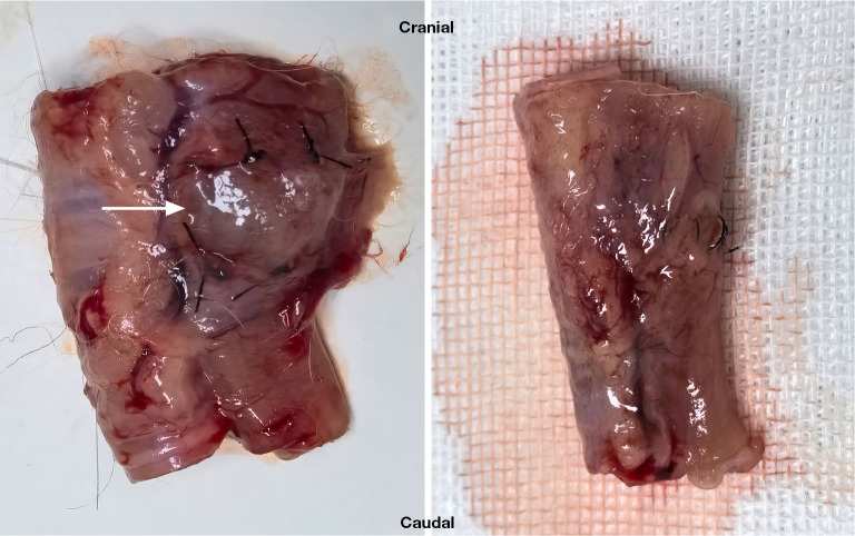
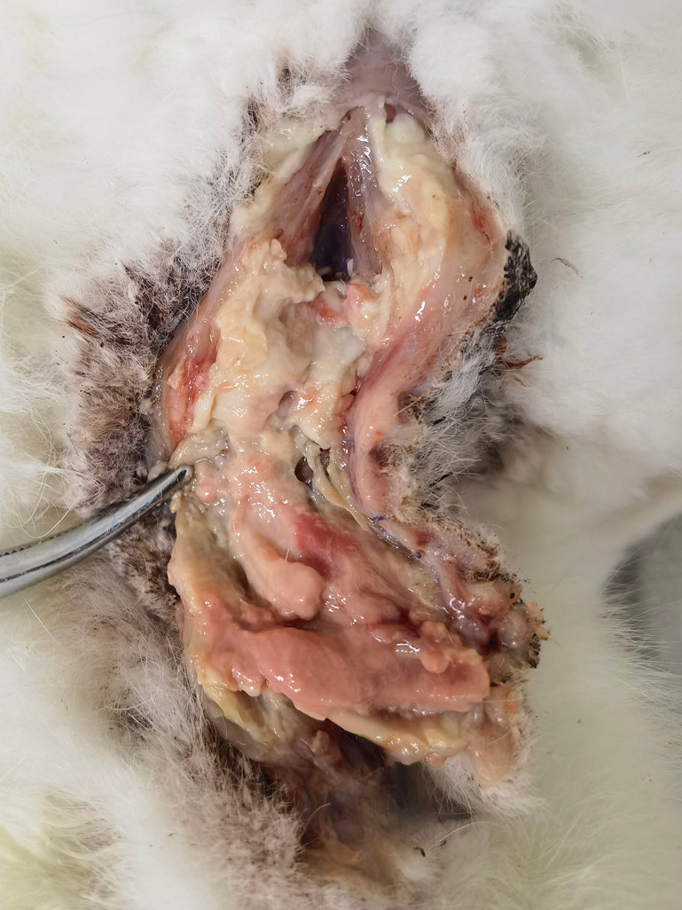
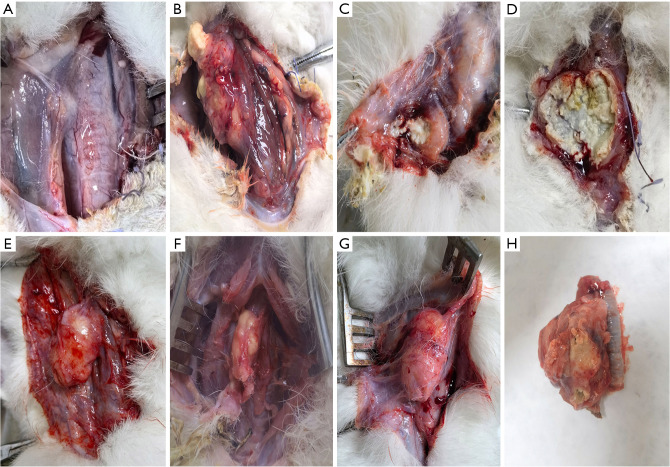
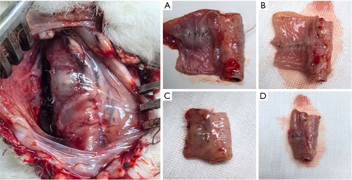
Forty large, healthy adult New Zealand white rabbits were randomized into the experimental group (n=20) and the control group (n=20). An animal model of cervical esophageal anastomosis was established using the esophagectomy anastomosis method. The esophageal anastomoses of the rabbits in the experimental group were completely covered with hydrogel biomaterial patches loaded with basic fibroblast growth factor (bFGF) intraoperatively. Four weeks after surgery, the incidence of anastomotic fistula was observed, and tissue samples were obtained from the esophageal anastomosis site for histologic and immunofluorescent detection.
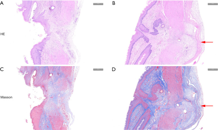
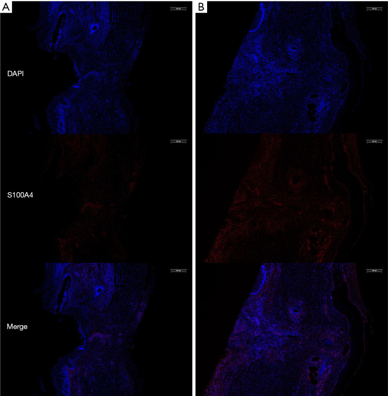
An animal model of cervical esophageal incision and anastomosis was successfully established in rabbits, and the incidence of anastomotic fistula in the experimental group was lower than that in the control group at the end of the experiment (1/18 versus 7/19; P<0.05). The esophageal anastomosis tensile test showed that the anastomosis in the experimental group had higher mechanical strength than that in the control group (6.49±0.17 versus 6.33±0.12 N; P<0.05). Histological examination of the anastomotic specimens showed that the tissue layers of the esophageal anastomoses in the experimental group were clear, and fibroblast proliferation and collagen fiber secretion were greater than those in the control group. Immunofluorescence showed that fibroblast proliferation was significantly increased in the perianastomotic tissues of the experimental group compared to that of the control group.
The application of hyaluronic acid methacrylate (HAMA) hydrogel biomaterial patches loaded with bFGF in esophageal surgery improved the mechanical strength of the esophageal anastomosis, facilitated fibroblast proliferation and collagen secretion, and could promote the growth of the tissues around the esophageal anastomosis. It may thus serve as a novel therapeutic approach for reducing the incidence of anastomotic fistula in esophageal cancer.
食管吻合口瘘的处理仍然是食管外科领域一项重大的临床挑战。随着材料科学的迅速发展,选择合适的生物材料来促进吻合口愈合并降低吻合口瘘的发生率已成为解决这些问题的一种有前景的策略。本研究的目的是探讨通过用合适的生物材料补片加强吻合来预防吻合口瘘。
将40只健康的成年新西兰大白兔随机分为实验组(n = 20)和对照组(n = 20)。采用食管切除吻合术建立颈段食管吻合动物模型。实验组兔的食管吻合口在术中用负载碱性成纤维细胞生长因子(bFGF)的水凝胶生物材料补片完全覆盖。术后4周,观察吻合口瘘的发生率,并从食管吻合部位获取组织样本进行组织学和免疫荧光检测。
成功在兔身上建立了颈段食管切开吻合动物模型,实验结束时实验组吻合口瘘的发生率低于对照组(1/18 vs 7/19;P<0.05)。食管吻合口拉伸试验表明,实验组的吻合口机械强度高于对照组(6.49±0.17 vs 6.33±0.12 N;P<0.05)。吻合口标本的组织学检查显示,实验组食管吻合口的组织层次清晰,成纤维细胞增殖和胶原纤维分泌多于对照组。免疫荧光显示,与对照组相比,实验组吻合口周围组织中的成纤维细胞增殖明显增加。
在食管手术中应用负载bFGF的甲基丙烯酸透明质酸(HAMA)水凝胶生物材料补片可提高食管吻合口的机械强度,促进成纤维细胞增殖和胶原分泌,并可促进食管吻合口周围组织的生长。因此,它可能作为一种新的治疗方法来降低食管癌吻合口瘘的发生率。