Steffens David C, Wang Lihong, Manning Kevin J, Pearlson Godfrey D
Department of Psychiatry (DCS, LW, KJM), University of Connecticut School of Medicine, Farmington CT; Departments of Psychiatry and Neuroscience (GDP), Yale University School of Medicine, New Haven CT; and the Olin Neuropsychiatry Research Center (GDP), Institute of Living, Hartford, CT.
Am J Geriatr Psychiatry Open Sci Educ Pract. 2025 Jun;6:1-14. doi: 10.1016/j.osep.2025.01.003. Epub 2025 Feb 7.
As alcohol use is common among older depressives, we assessed structural brain changes over 2 years and examined their association with changes in alcohol consumption.
Longitudinal cohort study.
Academic health center.
Adults aged 60 and older who met DSM criteria for a major depressive episode.
Participants were offered treatment with sertraline.
Participants completed structured interviews for reported alcohol consumption, had a clinical interview with a study psychiatrist, completed a cognitive battery at baseline and every twelve months, and underwent a 3T structural MRI as baseline and at 2-year follow-up. Volumetric brain changes were calculated.
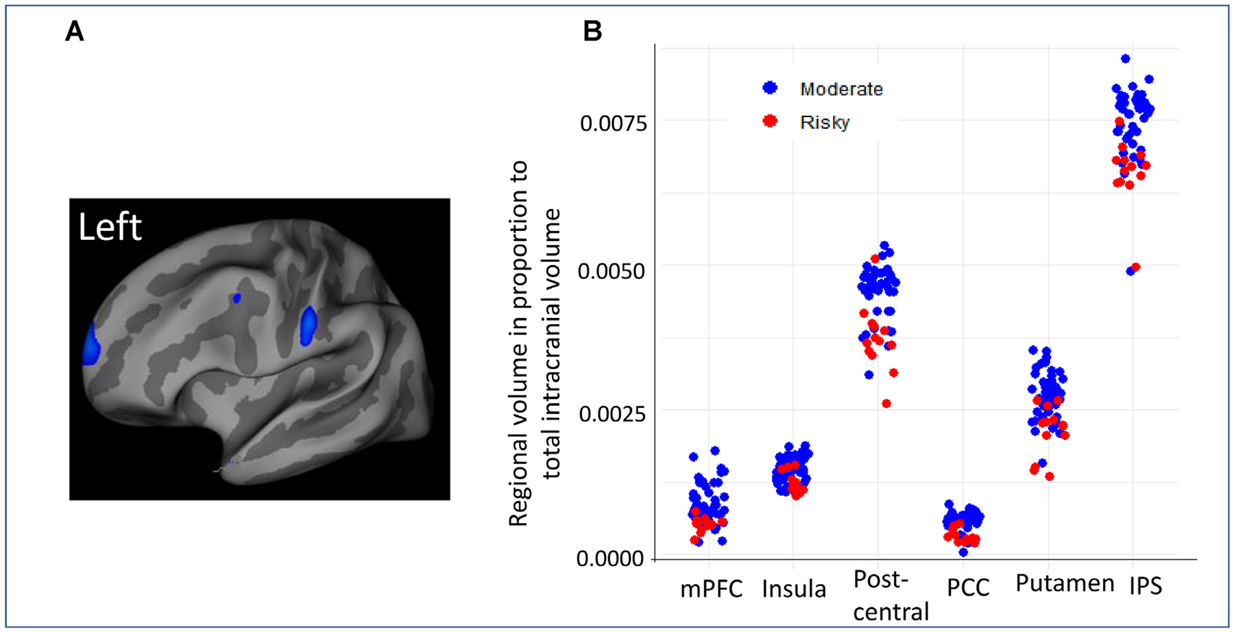
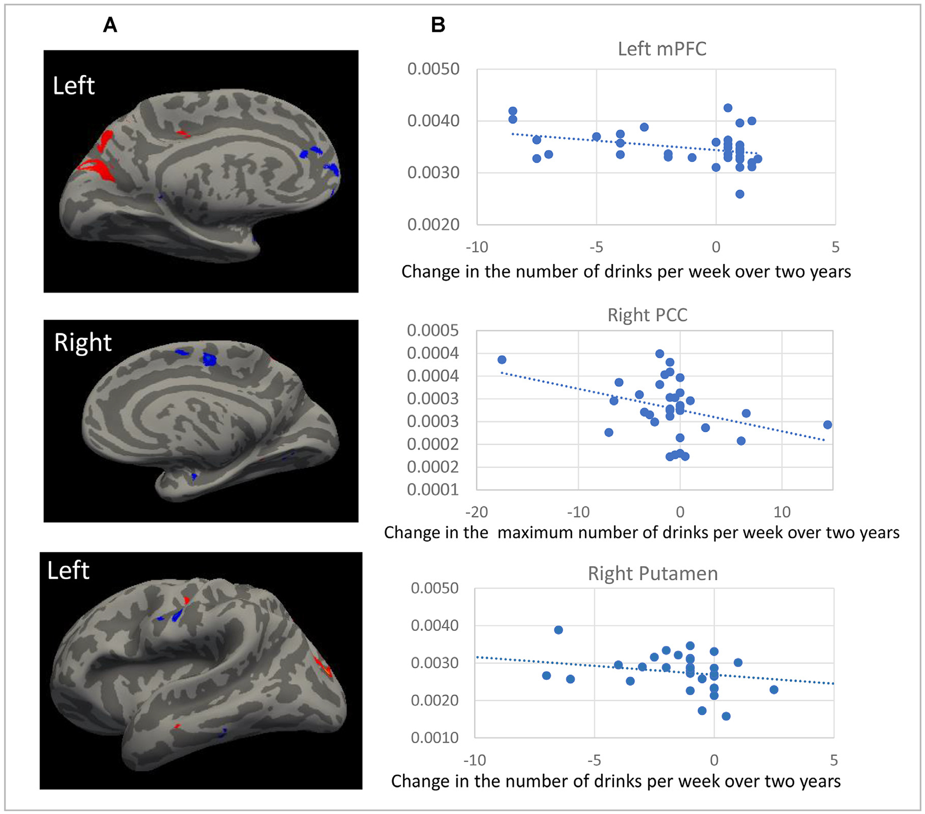
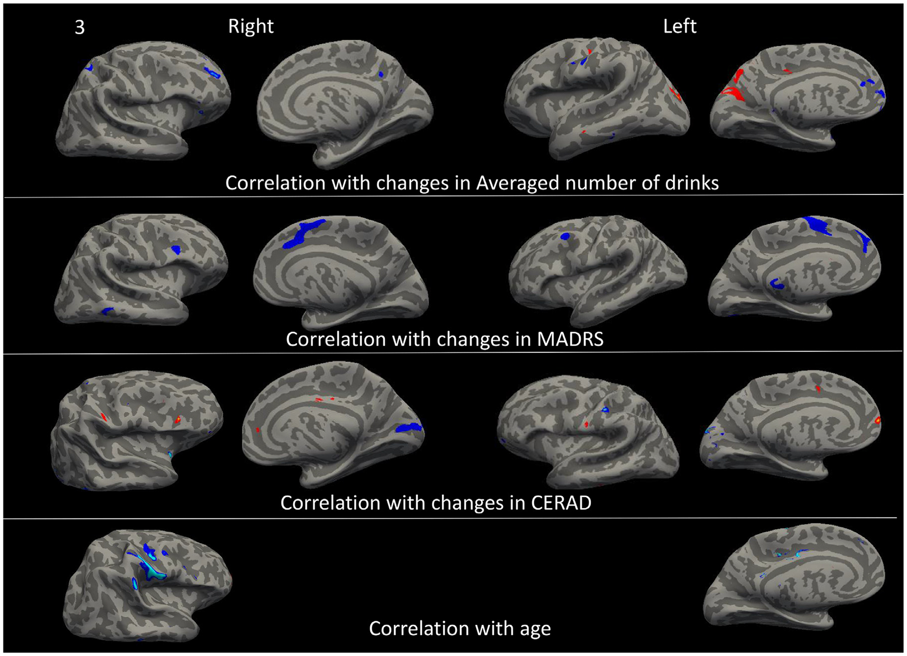
Among 58 participants, 45 were classified as moderate drinkers (≤7 drinks/week) and 13 as risky drinkers (>7 drinks/week). Compared with moderate drinkers, risky drinkers at baseline had significantly thinner cortical thickness and smaller volume in several frontal cortical regions, posterior cingulate, postcentral cortices, right insula, right putamen, and right inferior parietal sulcus. Annualized change in cortical thickness and volume correlated negatively with changes in the average number of drinks per week. Decreased depression severity, increased cognitive function score, and decreased alcohol consumption over the 2-year follow-up were each associated with annualized volumetric changes in specific common regions.
These MRI findings demonstrate the adverse impact of alcohol use in older adults on the fronto-striatal circuit. They highlight the need for careful screening and treatment referral for risky alcohol use among older adults with depression.
由于饮酒在老年抑郁症患者中很常见,我们评估了两年内大脑结构的变化,并研究了这些变化与饮酒量变化之间的关联。
纵向队列研究。
学术健康中心。
符合重度抑郁发作DSM标准的60岁及以上成年人。
为参与者提供舍曲林治疗。
参与者完成关于报告饮酒量的结构化访谈,与研究精神科医生进行临床访谈,在基线时和每十二个月完成一次认知测试,并在基线时和2年随访时接受3T结构MRI检查。计算大脑体积变化。
在58名参与者中,45名被归类为适度饮酒者(每周饮酒量≤7杯),13名被归类为高危饮酒者(每周饮酒量>7杯)。与适度饮酒者相比,基线时的高危饮酒者在几个额叶皮质区域、后扣带回、中央后皮质、右侧岛叶、右侧壳核和右侧顶下沟的皮质厚度明显更薄,体积更小。皮质厚度和体积的年化变化与每周平均饮酒量的变化呈负相关。在2年的随访中,抑郁严重程度降低、认知功能评分提高和饮酒量减少均与特定常见区域的年化体积变化相关。
这些MRI结果表明老年人饮酒对额纹状体回路有不利影响。它们强调了对患有抑郁症的老年人进行危险饮酒的仔细筛查和治疗转诊的必要性。