Lewis Josiah B, Fields Melanie E, Binkley Michael M, Zhou Anita, Mirro Amy, Ouyang Amy, Gupta Niket, Chen Yasheng, Fellah Slim, Smith Alyssa E, Dedkov Igor, Hulbert Monica L, Ford Andria L, An Hongyu, Lee Jin-Moo, Goyal Manu S, Guilliams Kristin P
Department of Neurology, Washington University School of Medicine, 660 S. Euclid Ave., St. Louis, MO, 63110, USA.
Department of Pediatrics, Washington University School of Medicine, 660 S. Euclid Ave., St. Louis, MO, 63110, USA.
Neuroimage Rep. 2025 Jun;5(2). doi: 10.1016/j.ynirp.2025.100265. Epub 2025 May 9.
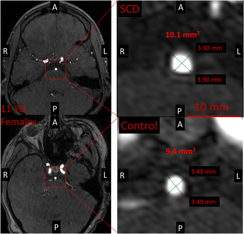
Children with sickle cell disease (SCD) may develop large vessel narrowing, but studies suggest vessels may also be enlarged, possibly related to increased cerebral blood flow (CBF). We used MRI to investigate whether the cross-sectional total inflow vessel luminal area (TIVLA) proximal to the circle of Willis (carotid arteries + basilar artery) would be increased in SCD compared to age- and sex-matched peers after adjusting for CBF. Across 36 children with SCD (19 female, median age 10.7 [8.0-14.5] years and 43 controls (26 female, median age 12.7 [9.2-18.2] years) matched by age ( = 0.13) and sex ( = 0.50), the median TIVLA in the SCD group (35.9 mm [30.7, 39.5]) was larger than controls (30.5 mm [27.8, 35.4], = 0.002). In a mixed model including age, sex, hemoglobin, CBF, SCD status, and an interaction between hemoglobin and SCD status, CBF (β = 0.11, CI 0.02-0.20, = 0.02), SCD (β = 28.02, CI 5.62-50.42, = 0.015), and the interaction between SCD and hemoglobin (β = -2.48, CI -4.49 to -0.47, = 0.018) were all significantly associated with increased TIVLA. Notably, TIVLA as a measure of arterial lumens is larger in children with SCD, even after adjusting for CBF in the mixed model. This implies disease-specific normative values may be needed to detect early vasculopathy.
患有镰状细胞病(SCD)的儿童可能会出现大血管狭窄,但研究表明血管也可能会扩张,这可能与脑血流量(CBF)增加有关。我们使用磁共振成像(MRI)来研究,在调整脑血流量后,与年龄和性别匹配的同龄人相比,患有SCD的儿童 Willis 环近端(颈动脉+基底动脉)的横断面总流入血管腔面积(TIVLA)是否会增加。在36名患有SCD的儿童(19名女性,中位年龄10.7[8.0 - 14.5]岁)和43名对照组儿童(26名女性,中位年龄12.7[9.2 - 18.2]岁)中,两组在年龄(P = 0.13)和性别(P = 0.50)上相匹配,SCD组的TIVLA中位数为35.9平方毫米[30.7, 39.5],大于对照组(30.5平方毫米[27.8, 35.4],P = 0.002)。在一个包含年龄、性别、血红蛋白、脑血流量、SCD状态以及血红蛋白与SCD状态之间相互作用的混合模型中,脑血流量(β = 0.11,可信区间0.02 - 0.20,P = 0.02)、SCD(β = 28.02,可信区间5.62 - 50.42,P = 0.015)以及SCD与血红蛋白之间的相互作用(β = -2.48,可信区间 - 4.49至 - 0.47,P = 0.018)均与TIVLA增加显著相关。值得注意的是,即使在混合模型中调整了脑血流量,作为动脉管腔测量指标的TIVLA在患有SCD的儿童中仍更大。这意味着可能需要特定疾病的标准值来检测早期血管病变。