Friedrich Philip, Grubitzsch Hanna, Wolf Benjamin, Eichholz Hannah M, Tutmarc Cary, Gottheil Pablo, Sauer Frank, Cornelis Alissa, Wegscheider Anne-Sophie, Aktas Bahriye, Käs Josef A, Stepan Holger
Peter Debye Institute for Soft Matter Physics, Leipzig University, 04103, Leipzig, Germany.
Department of Obstetrics, University Hospital Leipzig, 04103, Leipzig, Germany.
Sci Rep. 2025 Jun 20;15(1):20132. doi: 10.1038/s41598-025-04752-4.
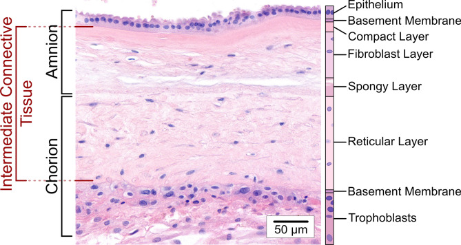
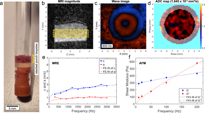
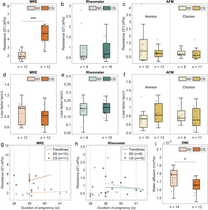
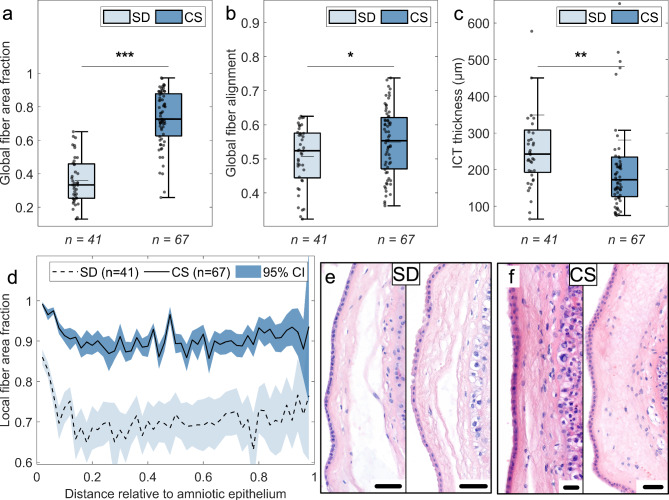
Rupture of fetal membranes and subsequent full-term birth are prerequisites for neonatal health, and a preterm rupture can lead to life-threatening complications. Our study determines the mechanical properties of term fetal membranes to identify perinatal structural changes by a unique biophysical multiscale approach, including atomic force microscopy, shear rheology, tabletop magnetic resonance elastography (MRE), and high-resolution optical microscopy. Fetal membranes from term spontaneous vaginal deliveries were compared to those from primary cesarean sections, used as a control group for pre-labor membranes. Spontaneously delivered term fetal membranes are softer and easier to deform in MRE experiments (median stiffness: 1.9 kPa, IQR 1.6-2.4) compared to controls (4.7 kPa, IQR 3.8-5.6); p < 0.001) and show increased water diffusion (median: 1.78 × 10 mm/s, IQR: (1.65-1.84) × 10 vs. 1.66 × 10 mm/s, IQR (1.60-1.73) × 10; p = 0.047). Their intermediate connective tissue layer (i.e. the collagen-rich area enclosed by the amnion and chorion) exhibits less ordered fiber alignment (median order parameter: 0.52, IQR 0.44-0.58 vs. 0.55, IQR 0.47-0.62; p = 0.04) and a looser fiber structure, as indicated by a significantly lower fiber area fraction (median: 0.33, IQR 0.25-0.46 vs. 0.73, IQR 0.63-0.88; p < 0.001) compared to the control membranes. These layer-specific changes in both structure and viscoelasticity are evidence for the dominant role of the intermediate connective tissue in maintaining membrane stability and the onset of rupture. Our mechanical and histopathological findings highlight the potential of mechanics-based screening-methods to assess the risk of preterm rupture and preterm birth to reduce neonatal morbidity.
胎膜破裂及随后的足月分娩是新生儿健康的先决条件,而胎膜早破会导致危及生命的并发症。我们的研究通过独特的生物物理多尺度方法,包括原子力显微镜、剪切流变学、桌面磁共振弹性成像(MRE)和高分辨率光学显微镜,来确定足月胎膜的力学性能,以识别围产期的结构变化。将足月自然阴道分娩的胎膜与初次剖宫产的胎膜进行比较,后者用作临产前胎膜的对照组。与对照组(4.7 kPa,四分位间距3.8 - 5.6)相比,自然分娩的足月胎膜在MRE实验中更柔软,更容易变形(中位硬度:1.9 kPa,四分位间距1.6 - 2.4);p < 0.001),并且水扩散增加(中位数:1.78×10⁻⁹ mm²/s,四分位间距:(1.65 - 1.84)×10⁻⁹ 对比1.66×10⁻⁹ mm²/s,四分位间距(1.60 - 1.73)×10⁻⁹;p = 0.047)。其中间结缔组织层(即羊膜和绒毛膜所包围的富含胶原蛋白的区域)显示出纤维排列的有序性较低(中位有序参数:0.52,四分位间距0.44 - 0.58对比0.55,四分位间距0.47 - 0.62;p = 0.04),并且纤维结构更松散,这表现为与对照膜相比,纤维面积分数显著更低(中位数:0.33,四分位间距0.25 - 0.46对比0.73,四分位间距0.63 - 0.88;p < 0.001)。这些结构和粘弹性方面的层特异性变化证明了中间结缔组织在维持胎膜稳定性和破裂起始中起主导作用。我们的力学和组织病理学发现突出了基于力学的筛查方法在评估胎膜早破和早产风险以降低新生儿发病率方面的潜力。