Goss Mark A, Burton Alex T, Kraus Jonathan C, McGrady Linda M, Wang Mei
Medical College of Wisconsin, Waukesha, WI, USA.
Medical College of Wisconsin, and Marquette University, Milwaukee, WI, USA.
Foot Ankle Orthop. 2025 Jun 19;10(2):24730114251342243. doi: 10.1177/24730114251342243. eCollection 2025 Apr.
Use of syndesmotic suture button fixation has gained in popularity for treating an injury to the tibiofibular syndesmosis. This biomechanical study used a cadaveric model to simulate in vivo loading conditions to assess the impact of the placement of a syndesmotic stabilization construct using a suture button device.
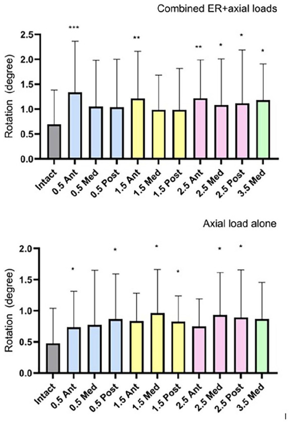
Biomechanical fixation stability with suture button device (TightRope; Arthrex, Naples, FL) placed at 4 distances from the tibiotalar joint line (0.5, 1.5, 2.5, and 3.5 cm) and 3 trajectories (anterior, medial, and posterior) were studied using cadaveric lower extremities with created syndesmotic injuries. Nondestructive testing was conducted on a biaxial servo-hydraulic load frame. The load application consisted of 2 portions: (1) axial compression simulating weightbearing and (2) external rotation of the ankle (up to 12 degrees and under 7.5 Nm) around the long axis of the tibia combined with weightbearing. Fibular motion and syndesmotic widening were tracked using motion analysis to quantify stability.
Fixation placed at 0.5 or 1.5 cm from the joint line in medial or posterior trajectories resulted in the lowest increases in fibular rotation under loading. More proximal or anterior placements led to increased fibular motion and decreased rotational stability. Ankle width changes were minimal in most groups, although slightly increased widening occurred at proximal and anterior placements.
Placement of the syndesmotic suture button fixation 0.5-1.5 cm of the joint line in medial or posterior orientations provides the most rotationally stable fixation in a cadaveric model. These findings support flexibility in syndesmotic suture button fixation positioning when hardware constraints limit ideal placement.
下胫腓联合缝线纽扣固定术在治疗胫腓下联合损伤中越来越受欢迎。本生物力学研究使用尸体模型模拟体内负荷条件,以评估使用缝线纽扣装置进行下胫腓联合稳定结构放置的影响。
使用造成下胫腓联合损伤的尸体下肢,研究将缝线纽扣装置(TightRope;Arthrex,那不勒斯,佛罗里达州)放置在距胫距关节线4个距离(0.5、1.5、2.5和3.5厘米)和3种轨迹(前、中、后)时的生物力学固定稳定性。在双轴伺服液压加载框架上进行无损测试。加载分为两部分:(1)模拟负重的轴向压缩;(2)踝关节围绕胫骨长轴的外旋(最大12度且扭矩低于7.5牛米)并结合负重。使用运动分析跟踪腓骨运动和下胫腓联合增宽,以量化稳定性。
在内侧或后侧轨迹中,距关节线0.5或1.5厘米处的固定在加载时腓骨旋转增加最少。更靠近近端或前方的放置会导致腓骨运动增加和旋转稳定性降低。大多数组的踝关节宽度变化最小,尽管在近端和前方放置时增宽略有增加。
在下胫腓联合缝线纽扣固定术中,在内侧或后侧方向距关节线0.5 - 1.5厘米处放置提供了尸体模型中最稳定的旋转固定。当硬件限制限制理想放置时,这些发现支持下胫腓联合缝线纽扣固定定位的灵活性。