• 文献检索
  • 文档翻译
  • 深度研究
  • 学术资讯
  • Suppr Zotero 插件Zotero 插件
  • 邀请有礼
  • 套餐&价格
  • 历史记录
应用&插件
Suppr Zotero 插件Zotero 插件浏览器插件Mac 客户端Windows 客户端微信小程序
定价
高级版会员购买积分包购买API积分包
服务
文献检索文档翻译深度研究API 文档MCP 服务
关于我们
关于 Suppr公司介绍联系我们用户协议隐私条款
关注我们

Suppr 超能文献

核心技术专利:CN118964589B侵权必究
粤ICP备2023148730 号-1Suppr @ 2026

文献检索

告别复杂PubMed语法,用中文像聊天一样搜索,搜遍4000万医学文献。AI智能推荐,让科研检索更轻松。

立即免费搜索

文件翻译

保留排版,准确专业,支持PDF/Word/PPT等文件格式,支持 12+语言互译。

免费翻译文档

深度研究

AI帮你快速写综述,25分钟生成高质量综述,智能提取关键信息,辅助科研写作。

立即免费体验

多模态超声检查在BI-RADS 4类乳腺良恶性病变鉴别诊断中的应用

The application of multimodal ultrasound examination in the differential diagnosis of benign and malignant breast lesions of BI-RADS category 4.

作者信息

Li Chunling, Wu Mumin, Haiqing Huang

机构信息

Cancer Hospital, College of Medicine, Shantou University, Shantou, China.

出版信息

Front Med (Lausanne). 2025 Jun 9;12:1596100. doi: 10.3389/fmed.2025.1596100. eCollection 2025.

DOI:10.3389/fmed.2025.1596100
PMID:40552183
原文链接:https://pmc.ncbi.nlm.nih.gov/articles/PMC12183244/
Abstract

OBJECTIVES

The purpose of this study was to the diagnostic value of conventional ultrasound (US), contrast-enhanced ultrasound (CEUS) and shear wave elastography (SWE) for identifying benign and malignant BI-RADS 4 breast lesions.

MATERIALS AND METHODS

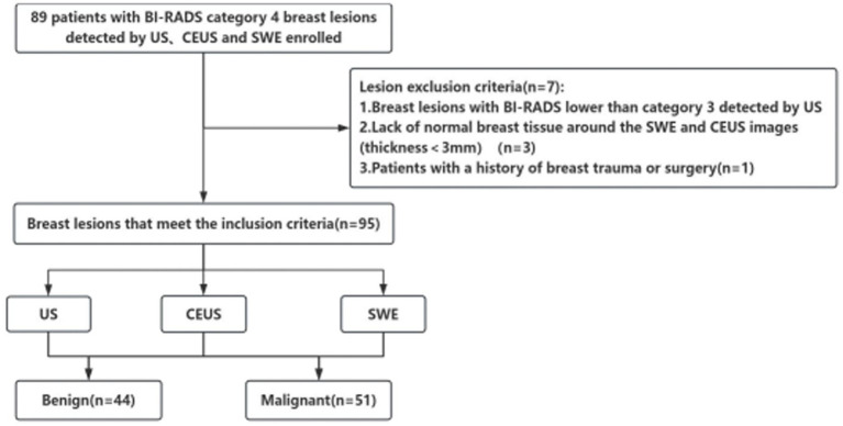
From February 2022 to November 2024, 95 patients aged 20 to 90 years with breast diseases, all of whom were female, were included. These lesions were diagnosed as BI-RADS 4 breast lesions by conventional ultrasound. All lesions were pathologically confirmed by surgical resection or tissue biopsy, and they were further evaluated by CEUS and SWE. The sensitivity, specificity, positive predictive value (PPV), negative predictive value (NPV), and accuracy of US, CEUS, and SWE were statistically analyzed, and ROC curves were generated. The diagnostic efficacy of US, US + SWE, US + CEUS, and US + CEUS + SWE were subsequently compared, with the pathology results used as the reference standard.

RESULTS

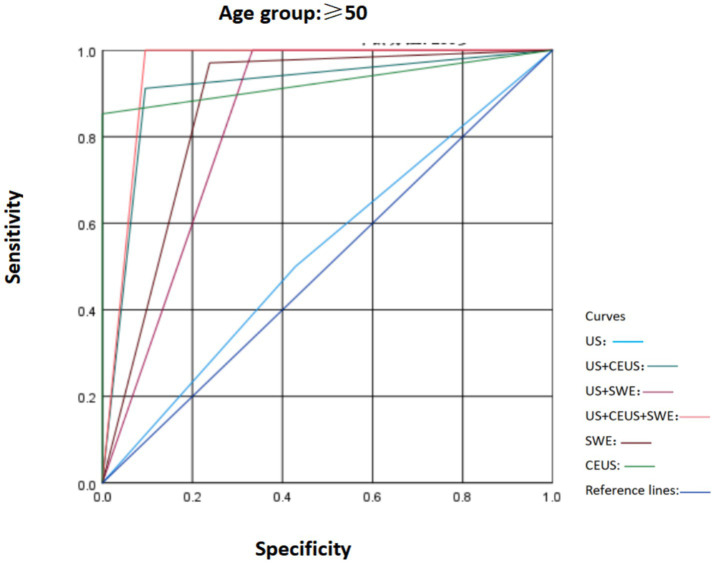
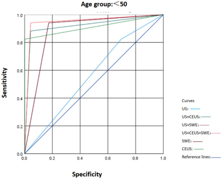
(1) Among the 95 BI-RADS 4 lesions, 44 (46.31%) were benign, and 51 (53.69%) were malignant. The sensitivity, specificity, PPV, NPV and accuracy of the BI-RADS classification via conventional US were 86.3, 72.7, 78.6, 82.1 and 80.0%, respectively. (2) The sensitivity, specificity, PPV, NPV, and accuracy of US combined with SWE in the diagnosis of breast nodules were 96.1, 79.5, 84.5, 94.6, and 88.4%, respectively. (3) The sensitivity, specificity, PPV, NPV, and accuracy of US combined with CEUS in the diagnosis of breast nodules were 84.3, 86.4, 87.8, 82.6, and 85.3%, respectively. (4) The areas under the ROC curve (AUCs) of US, US + SWE, and US + CEUS were 0.795, 0.877, and 0.917, respectively. Statistical methods were used to evaluate the US + CEUS + SWE method, and the results indicated excellent diagnostic performance. The AUC was 0.946, while the sensitivity, specificity, PPV, NPV, and accuracy were 90.7, 93.2, 94.2, 95.3, and 94.7%, respectively. In this this study, the AUCs of US, SWE, and CEUS were compared, and the results revealed that both SWE and CEUS could increase the AUC for breast lesion diagnosis with good diagnostic performance. These methods can increase the sensitivity, specificity and accuracy of the US examination when combined with conventional US. Moreover, the diagnostic performance for breast lesions was highest with the combined application of the three modalities, with a diagnostic AUC that was significantly higher than those of US alone, US + SWE and US + CEUS. The differences were significant ( < 0.05).

CONCLUSION

(1) CEUS and SWE provide diagnostic information about the microvascular perfusion and tissue stiffness of lesions, respectively, which can assist in the differentiation of benign from malignant breast tumors by conventional US and improve the sensitivity, specificity and accuracy of diagnosis, especially for US BI-RADS 4a breast lesions. (2) The combined use of CEUS and SWE in conventional US enhance the overall diagnostic performance with respect to breast lesions, with the best sensitivity and specificity and the highest diagnostic efficacy. The use of US + CEUS + SWE is beneficial for further differentiating benign and malignant breast lesions according to the US BI-RADS 4, thereby reducing unnecessary biopsies or surgeries.

https://cdn.ncbi.nlm.nih.gov/pmc/blobs/4356/12183244/7eef7d4d0ab8/fmed-12-1596100-g006.jpg
https://cdn.ncbi.nlm.nih.gov/pmc/blobs/4356/12183244/de866ae9f818/fmed-12-1596100-g001.jpg
https://cdn.ncbi.nlm.nih.gov/pmc/blobs/4356/12183244/4e3dd9680327/fmed-12-1596100-g002.jpg
https://cdn.ncbi.nlm.nih.gov/pmc/blobs/4356/12183244/8e767437d2fb/fmed-12-1596100-g003.jpg
https://cdn.ncbi.nlm.nih.gov/pmc/blobs/4356/12183244/901b8906e921/fmed-12-1596100-g004.jpg
https://cdn.ncbi.nlm.nih.gov/pmc/blobs/4356/12183244/44841150a95e/fmed-12-1596100-g005.jpg
https://cdn.ncbi.nlm.nih.gov/pmc/blobs/4356/12183244/7eef7d4d0ab8/fmed-12-1596100-g006.jpg
摘要

目的

本研究旨在探讨常规超声(US)、超声造影(CEUS)和剪切波弹性成像(SWE)对鉴别乳腺影像报告和数据系统(BI-RADS)4类乳腺病变良恶性的诊断价值。

材料与方法

纳入2022年2月至2024年11月95例年龄20至90岁的乳腺疾病患者,均为女性。这些病变经常规超声诊断为BI-RADS 4类乳腺病变。所有病变均经手术切除或组织活检病理证实,并进一步行CEUS和SWE评估。对US、CEUS和SWE的敏感性、特异性、阳性预测值(PPV)、阴性预测值(NPV)和准确性进行统计学分析,并绘制ROC曲线。随后以病理结果为参考标准,比较US、US + SWE、US + CEUS和US + CEUS + SWE的诊断效能。

结果

(1)95例BI-RADS 4类病变中,良性44例(46.31%),恶性51例(53.69%)。常规US的BI-RADS分类的敏感性、特异性、PPV、NPV和准确性分别为86.3%、72.7%、78.6%、82.1%和80.0%。(2)US联合SWE诊断乳腺结节的敏感性、特异性、PPV、NPV和准确性分别为96.1%、79.5%、84.5%、94.6%和88.4%。(3)US联合CEUS诊断乳腺结节的敏感性、特异性、PPV、NPV和准确性分别为84.3%、86.4%、87.8%、82.6%和85.3%。(4)US、US + SWE和US + CEUS的ROC曲线下面积(AUC)分别为0.795、0.877和0.917。采用统计方法评估US + CEUS + SWE方法,结果显示其具有优异的诊断性能。AUC为0.946,敏感性、特异性、PPV、NPV和准确性分别为90.7%、93.2%、94.2%、95.3%和94.7%。本研究比较了US、SWE和CEUS的AUC,结果显示SWE和CEUS均可提高乳腺病变诊断的AUC,诊断性能良好。这些方法与常规US联合使用时可提高US检查的敏感性、特异性和准确性。此外,三种模式联合应用对乳腺病变的诊断性能最高,诊断AUC显著高于单独使用US、US + SWE和US + CEUS。差异有统计学意义(P < 0.05)。

结论

(1)CEUS和SWE分别提供病变的微血管灌注和组织硬度的诊断信息,可辅助常规US鉴别乳腺肿瘤的良恶性,提高诊断的敏感性、特异性和准确性,尤其对于US BI-RADS 4a类乳腺病变。(2)在常规US中联合使用CEUS和SWE可提高乳腺病变的整体诊断性能,具有最佳的敏感性和特异性以及最高的诊断效能。使用US + CEUS + SWE有利于根据US BI-RADS 4进一步鉴别乳腺病变的良恶性,从而减少不必要的活检或手术。

相似文献

1
The application of multimodal ultrasound examination in the differential diagnosis of benign and malignant breast lesions of BI-RADS category 4.多模态超声检查在BI-RADS 4类乳腺良恶性病变鉴别诊断中的应用
Front Med (Lausanne). 2025 Jun 9;12:1596100. doi: 10.3389/fmed.2025.1596100. eCollection 2025.
2
Using a Large Language Model for Breast Imaging Reporting and Data System Classification and Malignancy Prediction to Enhance Breast Ultrasound Diagnosis: Retrospective Study.使用大语言模型进行乳腺影像报告和数据系统分类及恶性肿瘤预测以增强乳腺超声诊断:回顾性研究
JMIR Med Inform. 2025 Jun 11;13:e70924. doi: 10.2196/70924.
3
BI-RADS 4 breast lesions: could multi-mode ultrasound be helpful for their diagnosis?乳腺影像报告和数据系统(BI-RADS)4类乳腺病变:多模式超声对其诊断有帮助吗?
Gland Surg. 2019 Jun;8(3):258-270. doi: 10.21037/gs.2019.05.01.
4
Signs and symptoms to determine if a patient presenting in primary care or hospital outpatient settings has COVID-19.在基层医疗机构或医院门诊环境中,如果患者出现以下症状和体征,可判断其是否患有 COVID-19。
Cochrane Database Syst Rev. 2022 May 20;5(5):CD013665. doi: 10.1002/14651858.CD013665.pub3.
5
Transabdominal ultrasound and endoscopic ultrasound for diagnosis of gallbladder polyps.经腹超声和内镜超声用于胆囊息肉的诊断。
Cochrane Database Syst Rev. 2018 Aug 15;8(8):CD012233. doi: 10.1002/14651858.CD012233.pub2.
6
Contrast-enhanced ultrasound for the diagnosis of hepatocellular carcinoma in adults with chronic liver disease.对比增强超声在慢性肝病成人肝细胞癌诊断中的应用。
Cochrane Database Syst Rev. 2022 Sep 2;9(9):CD013483. doi: 10.1002/14651858.CD013483.pub2.
7
Application Value of Deep Learning-Based AI Model in the Classification of Breast Nodules.基于深度学习的人工智能模型在乳腺结节分类中的应用价值
Br J Hosp Med (Lond). 2025 Jun 25;86(6):1-19. doi: 10.12968/hmed.2025.0078. Epub 2025 Jun 15.
8
Three-dimensional saline infusion sonography compared to two-dimensional saline infusion sonography for the diagnosis of focal intracavitary lesions.三维盐水灌注超声与二维盐水灌注超声在诊断腔内局灶性病变中的比较。
Cochrane Database Syst Rev. 2017 May 5;5(5):CD011126. doi: 10.1002/14651858.CD011126.pub2.
9
Efficacy of shear wave elastography in measuring placental stiffness for pre-eclampsia screening and diagnosis: A systematic review and meta-analysis.剪切波弹性成像测量胎盘硬度用于子痫前期筛查和诊断的有效性:一项系统评价和荟萃分析
Int J Gynaecol Obstet. 2025 Jun 23. doi: 10.1002/ijgo.70293.
10
Magnetic resonance perfusion for differentiating low-grade from high-grade gliomas at first presentation.首次就诊时磁共振灌注成像用于鉴别低级别与高级别胶质瘤
Cochrane Database Syst Rev. 2018 Jan 22;1(1):CD011551. doi: 10.1002/14651858.CD011551.pub2.

本文引用的文献

1
Systematic Meta-Analysis of Computer-Aided Detection of Breast Cancer Using Hyperspectral Imaging.使用高光谱成像技术对乳腺癌计算机辅助检测的系统Meta分析。
Bioengineering (Basel). 2024 Oct 24;11(11):1060. doi: 10.3390/bioengineering11111060.
2
Ultrasound as a Method for Early Diagnosis of Breast Pathology.超声作为乳腺疾病早期诊断的一种方法。
J Pers Med. 2023 Jul 18;13(7):1156. doi: 10.3390/jpm13071156.
3
Feasibility of Diffusion Tensor Imaging for Decreasing Biopsy Rates in Breast Imaging: Interim Analysis of a Prospective Study.
扩散张量成像在降低乳腺成像活检率中的可行性:一项前瞻性研究的中期分析
Diagnostics (Basel). 2023 Jun 30;13(13):2226. doi: 10.3390/diagnostics13132226.
4
Breast Imaging Reporting and Data System and Contrast Enhancement Mammography: Lesion Conspicuity Likelihood of Malignancy and Relationship With Breast Tumor Receptor Status.乳腺影像报告和数据系统与对比增强乳腺摄影:病灶显著性恶性可能性及其与乳腺癌受体状态的关系。
Acad Radiol. 2023 Oct;30(10):2243-2251. doi: 10.1016/j.acra.2023.02.008. Epub 2023 Mar 8.
5
Global Cancer Statistics 2020: GLOBOCAN Estimates of Incidence and Mortality Worldwide for 36 Cancers in 185 Countries.《全球癌症统计数据 2020:全球 185 个国家和地区 36 种癌症的发病率和死亡率估计》。
CA Cancer J Clin. 2021 May;71(3):209-249. doi: 10.3322/caac.21660. Epub 2021 Feb 4.
6
The role of tissue elasticity in the differential diagnosis of benign and malignant breast lesions using shear wave elastography.剪切波弹性成像技术在鉴别良恶性乳腺病变中的组织弹性作用。
BMC Cancer. 2020 Sep 29;20(1):930. doi: 10.1186/s12885-020-07423-x.
7
Value of quantitative sound touch elastography of tissues around breast lesions in the evaluation of malignancy.乳腺病变周围组织声触诊弹性定量技术评估恶性价值。
Clin Radiol. 2021 Jan;76(1):79.e21-79.e28. doi: 10.1016/j.crad.2020.08.016. Epub 2020 Sep 15.
8
Multimodal Ultrasound Imaging in Breast Imaging-Reporting and Data System 4 Breast Lesions: A Prediction Model for Malignancy.多模态超声成像在乳腺影像报告和数据系统 4 类乳腺病变中的应用:恶性肿瘤预测模型。
Ultrasound Med Biol. 2020 Dec;46(12):3188-3199. doi: 10.1016/j.ultrasmedbio.2020.08.003. Epub 2020 Sep 4.
9
Diffusion-weighted MRI for Unenhanced Breast Cancer Screening.弥散加权 MRI 在乳腺癌筛查中的应用。
Radiology. 2019 Dec;293(3):504-520. doi: 10.1148/radiol.2019182789. Epub 2019 Oct 8.
10
Screening for Breast Cancer in Average-Risk Women: A Guidance Statement From the American College of Physicians.美国医师学院关于一般风险女性乳腺癌筛查的指南声明。
Ann Intern Med. 2019 Apr 16;170(8):547-560. doi: 10.7326/M18-2147. Epub 2019 Apr 9.