Figueroa Esteban G, Castillo Rodrigo L, Paz Adolfo A, Monsalves-Alvarez Matías, Salas-Pérez Francisca, Calle Ximena, Jiménez Tamara A, Herrera Emilio A, Gonzaléz-Candia Alejandro
Laboratory of Fetal Neuroprogramming, Institute of Health Sciences, Universidad de O'Higgins, Rancagua 3655000, Chile.
Institute of Health Sciences, Universidad de O'Higgins, Rancagua 3655000, Chile.
Antioxidants (Basel). 2025 Jun 15;14(6):736. doi: 10.3390/antiox14060736.
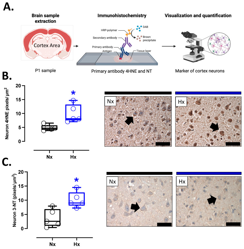
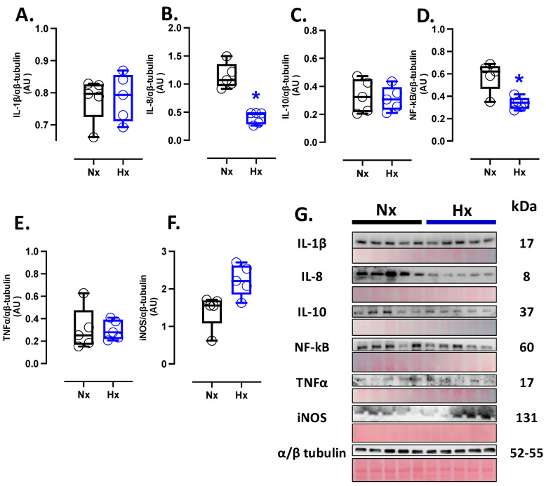
Gestational chronic hypoxia impacts prenatal development, leading to fetal growth restriction (FGR), defined as the fetus's failure to reach its genetic growth potential. Postnatal hypoxia in the cerebral tissue can induce a redox imbalance and mitochondrial dysfunction, consequently increasing neuronal death. However, these data cannot necessarily be extrapolated to prenatal hypoxia. In this regard, this study aims to describe the effect of gestational hypoxia on redox balance and apoptosis cell death mechanisms in the prefrontal cortex of guinea pigs. Ten Guinea pig () pregnant dams were utilized in this study; five gestated in normoxia (Nx; three newborn males, and two females) and five gestated under chronic hypobaric hypoxia (Hx; two newborn males, and three females). We monitored the pregnancies by ultrasound examinations from gestational days 20 to 65 (term ~ 70). At birth, pups were euthanized, and the fetal brain was collected for cellular redox measurement, mitochondrial enzyme expression, and apoptosis assay. Gestation under hypoxia induced an imbalance in the expression of anti- and pro-oxidant enzymes, resulting in increased oxidative stress. Additionally, a decrease in cytochrome I and III expression and neuronal density in the neonatal prefrontal cortex was observed. Finally, DNA fragmentation was increased by the TUNEL assay in the brain tissue of newborns gestated under chronic hypoxia. Our findings demonstrate the association of gestational hypoxia with oxidative stress and neuronal death in newborns, which may predispose to neuronal dysfunction in adulthood.
妊娠期慢性缺氧会影响产前发育,导致胎儿生长受限(FGR),即胎儿无法达到其遗传生长潜力。脑组织的产后缺氧可诱导氧化还原失衡和线粒体功能障碍,从而增加神经元死亡。然而,这些数据不一定能外推至产前缺氧情况。在这方面,本研究旨在描述妊娠期缺氧对豚鼠前额叶皮质氧化还原平衡和凋亡细胞死亡机制的影响。本研究使用了10只豚鼠怀孕母鼠;5只在常氧环境下妊娠(Nx;3只新生雄性和2只雌性),5只在慢性低压缺氧环境下妊娠(Hx;2只新生雄性和3只雌性)。我们从妊娠第20天至65天(足月约70天)通过超声检查监测妊娠情况。出生时,对幼崽实施安乐死,并收集胎儿大脑进行细胞氧化还原测量、线粒体酶表达和凋亡检测。缺氧环境下的妊娠导致抗氧化酶和促氧化酶表达失衡,从而增加氧化应激。此外,还观察到新生儿前额叶皮质中细胞色素I和III表达以及神经元密度降低。最后,通过TUNEL检测发现,慢性缺氧环境下妊娠的新生儿脑组织中的DNA片段化增加。我们的研究结果表明,妊娠期缺氧与新生儿氧化应激和神经元死亡有关,这可能会使成年后易患神经元功能障碍。