Hong Charles Chong-Hwa
Patuxent Institution, Correctional Mental Health Center-Jessup, Jessup, MD 20794, USA.
Department of Psychiatry and Behavioral Sciences, The Johns Hopkins Hospital, Baltimore, MD 21205, USA.
Brain Sci. 2025 May 26;15(6):574. doi: 10.3390/brainsci15060574.
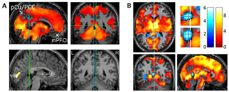
With positron emission tomography followed by functional magnetic resonance imaging (fMRI), we demonstrated that rapid eye movements (REMs) in sleep are saccades that scan dream imagery. The brain "sees" essentially the same way while awake and while dreaming in REM sleep. As expected, an event-related fMRI study (events = REMs) showed activation time-locked to REMs in sleep ("REM-locked" activation) in the oculomotor circuit that controls saccadic eye movements and visual attention. More crucially, the fMRI study provided a series of unexpected findings, including REM-locked multisensory integration. REMs in sleep index the processing of endogenous visual information and the hierarchical generation of dream imagery through multisensory integration. The neural processes concurrent with REMs overlap extensively with those reported to be atypical in autism spectrum disorder (ASD). Studies on ASD have shown atypical visual processing and multisensory integration, emerging early in infancy and subsequently developing into autistic symptoms. MRI studies of infants at high risk for ASD are typically conducted during natural sleep. Simply timing REMs may improve the accuracy of early detection and identify markers for stratification in heterogeneous ASD patients. REMs serve as a task-free probe useful for studying both infants and animals, who cannot comply with conventional visual activation tasks. Note that REM-probe studies would be easier to implement in early infancy because REM sleep, which is markedly preponderant in the last trimester of pregnancy, is still pronounced in early infancy. The brain may practice seeing the world during REM sleep in utero before birth. The REM-probe controls the level of attention across both the lifespan and typical-atypical neurodevelopment. Longitudinal REM-probe studies may elucidate how the brain develops the ability to "see" and how this goes awry in autism. REMs in sleep may allow a straightforward comparison of animal and human data. REM-probe studies of animal models of autism have great potential. This narrative review puts forth every reason to believe that employing REMs as a probe into the development of the visual brain will have far-reaching implications.
通过正电子发射断层扫描,随后进行功能磁共振成像(fMRI),我们证明了睡眠中的快速眼动(REM)是扫视运动,用于扫描梦境图像。大脑在清醒和快速眼动睡眠中做梦时的“看”的方式基本相同。正如预期的那样,一项事件相关的fMRI研究(事件=快速眼动)显示,在控制眼球扫视运动和视觉注意力的动眼神经回路中,睡眠中的快速眼动会出现时间锁定的激活(“快速眼动锁定”激活)。更关键的是,fMRI研究提供了一系列意想不到的发现,包括快速眼动锁定的多感官整合。睡眠中的快速眼动通过多感官整合来索引内源性视觉信息的处理和梦境图像的分层生成。与快速眼动同时发生的神经过程与自闭症谱系障碍(ASD)中报道的非典型神经过程广泛重叠。对自闭症谱系障碍的研究表明,早期婴儿期就出现了非典型的视觉处理和多感官整合,随后发展为自闭症症状。对自闭症高风险婴儿的MRI研究通常在自然睡眠期间进行。简单地对快速眼动进行计时可能会提高早期检测的准确性,并识别异质性自闭症谱系障碍患者的分层标志物。快速眼动可作为一种无需任务的探针,有助于研究婴儿和动物,因为他们无法完成传统的视觉激活任务。请注意,快速眼动探针研究在婴儿早期更容易实施,因为快速眼动睡眠在妊娠晚期明显占优势,在婴儿早期仍然很明显。大脑可能在出生前的子宫内快速眼动睡眠期间练习看世界。快速眼动探针在整个生命周期和典型-非典型神经发育过程中控制注意力水平。纵向快速眼动探针研究可能会阐明大脑如何发展“看”的能力,以及这种能力在自闭症中是如何出错的。睡眠中的快速眼动可能允许直接比较动物和人类数据。对自闭症动物模型的快速眼动探针研究具有巨大潜力。这篇叙述性综述提出了充分的理由相信,将快速眼动用作探究视觉大脑发育的探针将产生深远影响。