Majos Marcin, Klepaczko Artur, Szychowska Katarzyna, Stefanczyk Ludomir, Kurnatowska Ilona
Department of Normal and Clinical Anatomy, Medical University of Lodz, 90-419 Lodz, Poland.
I Department of Radiology and Diagnostic Imaging, Medical University of Lodz, 90-419 Lodz, Poland.
Biomedicines. 2025 Jun 4;13(6):1381. doi: 10.3390/biomedicines13061381.
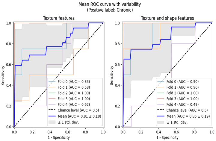
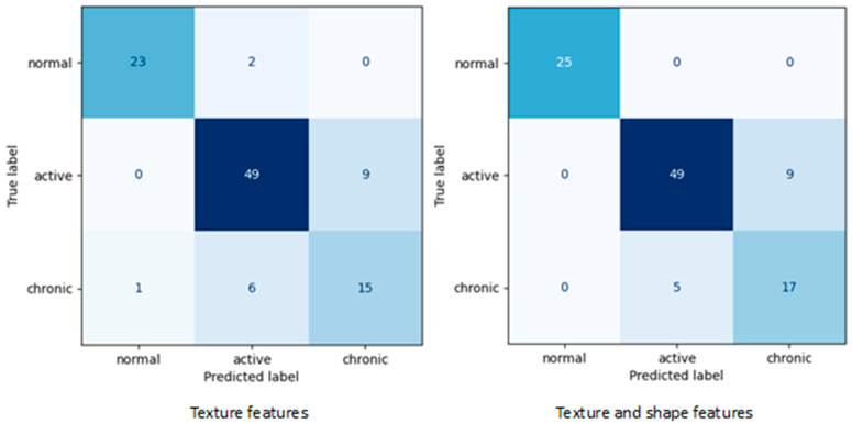
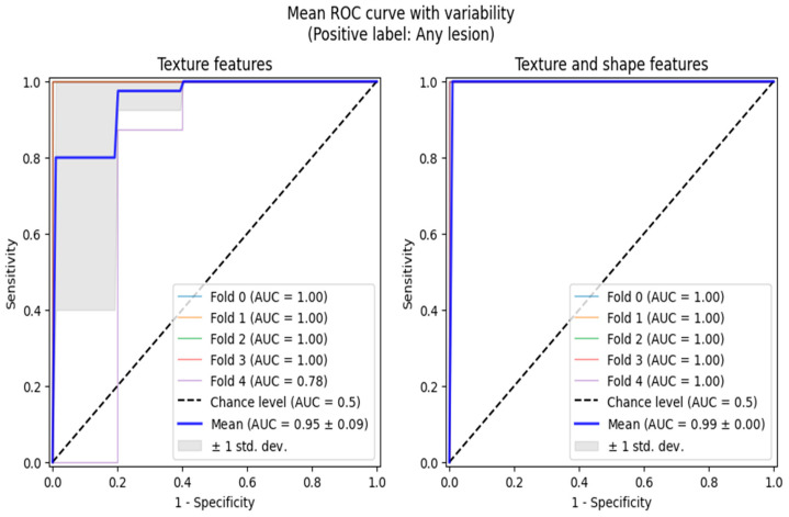
: The diagnostics of chronic kidney disease (CKD) consist of three basic groups of examinations: laboratory tests, radiological imaging and histopathological examinations. However, in the most severe clinical cases, where a fast, undisputed decision is required, histopathological tests are the only suitable option. Unfortunately, such tests require an invasive kidney biopsy, which is not possible in many patients. The aim of this study is to create an algorithm that can categorize CKD patients into active and non-active phases on the basis of MRI texture analysis and compare the results with histopathological examinations. : MRI examinations were performed on healthy volunteers (group 1, N = 14) and CKD patients who also received kidney biopsy. The histopathological examination was used to divide the patients into active phase CKD (group 2, N = 58) and non-active phase CKD (group 3, N = 22). The T2-weighted MRI images were analyzed using a Support Vector Machine (SVM) model created with qMazDa software, which was trained to classify images into the appropriate group of CKD activity. : The following evaluation metrics were calculated for the final SVM models corresponding to confusion matrices: for texture analysis-balanced accuracy 81.6%, sensitivity 68.2-92.0%, specificity 82.5-97.5% and precision 62.5-95.8%; for texture and shape analysis-balanced accuracy 87.3%, sensitivity 77.3-100.0%, specificity 87.5-100.0% and precision 65.4-100.0%. : Texture analysis of T2-weighted images associated with kidney shape features seems to be reliable method of assessing the state of ongoing CKD.
慢性肾脏病(CKD)的诊断包括三组基本检查:实验室检查、放射影像学检查和组织病理学检查。然而,在最严重的临床病例中,需要快速、无争议的诊断,组织病理学检查是唯一合适的选择。不幸的是,此类检查需要进行侵入性肾活检,而许多患者无法进行。本研究的目的是创建一种算法,该算法可以基于MRI纹理分析将CKD患者分为活动期和非活动期,并将结果与组织病理学检查进行比较。
对健康志愿者(第1组,N = 14)和也接受了肾活检的CKD患者进行了MRI检查。组织病理学检查用于将患者分为CKD活动期(第2组,N = 58)和非CKD活动期(第3组,N = 22)。使用qMazDa软件创建的支持向量机(SVM)模型分析T2加权MRI图像,该模型经过训练可将图像分类到适当的CKD活动组。
针对与混淆矩阵对应的最终SVM模型计算了以下评估指标:纹理分析——平衡准确率81.6% , 灵敏度68.2 - 92.0% , 特异性82.5 - 97.5% , 精确率62.5 - 95.8%;纹理和形状分析——平衡准确率87.3% , 灵敏度77.3 - 100.0% , 特异性87.5 - 100.0% , 精确率65.4 - 100.0%。
与肾脏形状特征相关的T2加权图像的纹理分析似乎是评估正在进行的CKD状态的可靠方法。