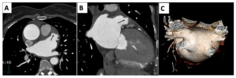
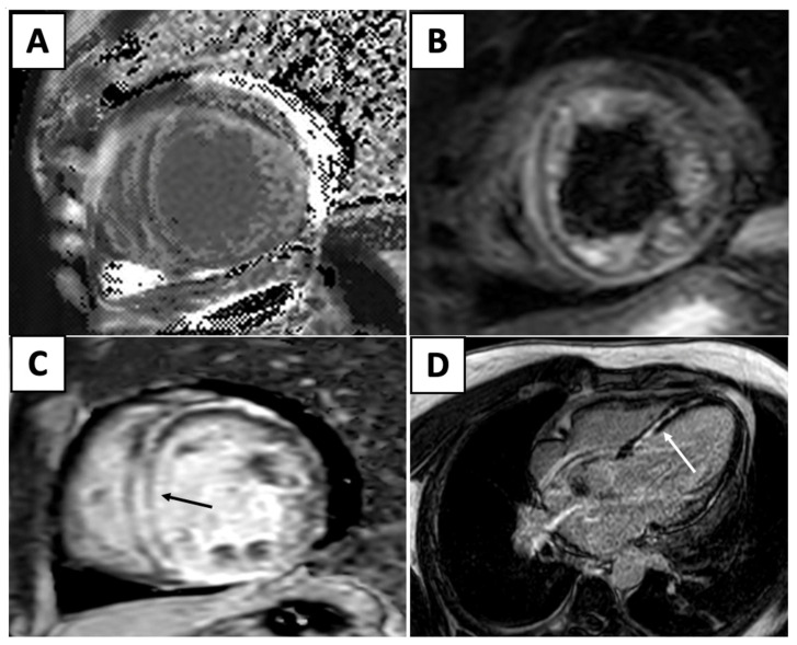
Park Christine M, Lerman Ben, Contreras Yametti Felipe, Garcia Mario, Slipczuk Leandro, Schenone Aldo L, Zhang Lili, Gongora Carlos A
Division of Cardiology, Montefiore Medical Center, Albert Einstein College of Medicine, Bronx, NY 10461, USA.
Department of Medicine, Montefiore Medical Center, Albert Einstein College of Medicine, Bronx, NY 10461, USA.
J Clin Med. 2025 Jun 18;14(12):4353. doi: 10.3390/jcm14124353.
Early detection and the rise of targeted cancer treatment have led to increased overall survival and decreased mortality among cancer patients. As the cancer survivor population ages, there is an increased risk for cardiovascular disease due to pre-existing comorbidities, deconditioning during therapy, or the natural progression of aging. Furthermore, with emerging oncologic therapies, there is an increased recognition of their potential cardiovascular toxicities. Indeed, heart disease is the leading cause of death in cancer survivors, which may reflect upon both the success of novel oncologic therapies and their potential cardiovascular toxicities. This recognition has driven the development of cardio-oncology, a multi-disciplinary field that involves collaboration between hematologists, oncologists, and cardiologists to screen, prevent, and manage cardiovascular disease in cancer patients and cancer survivors. The field focuses on early cardiovascular detection and prevention for these patients before, during, and after their oncologic treatment. As oncologic therapies evolve and our knowledge of short- and long-term adverse cardiovascular effects grows, it is critical for physicians to identify those at risk for increased morbidity and mortality, while also balancing the importance of their oncologic treatment plan. Multimodality cardiac imaging is the crux of the diagnosis and surveillance of these patients within cardio-oncology, and includes echocardiography, nuclear, computed tomography (CT), and cardiac magnetic resonance (CMR). Cardiac imaging is essential to establish the baseline function and assess various cardiotoxicities, including left ventricular dysfunction, heart failure, atherosclerosis, vascular injury, and arrhythmias. This review will discuss common oncologic therapies and their cardiotoxic profiles, the cardiac multimodality imaging modalities used in cardio-oncology, and the various approaches for the diagnosis and surveillance of this population.
早期检测和靶向癌症治疗的兴起提高了癌症患者的总体生存率并降低了死亡率。随着癌症幸存者群体的老龄化,由于先前存在的合并症、治疗期间的身体机能下降或衰老的自然进程,心血管疾病的风险增加。此外,随着新兴肿瘤治疗方法的出现,人们越来越认识到它们潜在的心血管毒性。事实上,心脏病是癌症幸存者的主要死因,这可能既反映了新型肿瘤治疗方法的成功,也反映了它们潜在的心血管毒性。这种认识推动了心脏肿瘤学的发展,这是一个多学科领域,涉及血液科医生、肿瘤内科医生和心脏病专家之间的合作,以筛查、预防和管理癌症患者和癌症幸存者的心血管疾病。该领域专注于在肿瘤治疗前、治疗期间和治疗后对这些患者进行早期心血管检测和预防。随着肿瘤治疗方法的不断发展以及我们对短期和长期心血管不良影响的认识不断增加,医生识别那些发病和死亡风险增加的患者至关重要,同时还要平衡肿瘤治疗计划的重要性。多模态心脏成像技术是心脏肿瘤学中这些患者诊断和监测的关键,包括超声心动图、核医学、计算机断层扫描(CT)和心脏磁共振成像(CMR)。心脏成像对于确定基线功能和评估各种心脏毒性至关重要,这些毒性包括左心室功能障碍、心力衰竭、动脉粥样硬化、血管损伤和心律失常。本综述将讨论常见的肿瘤治疗方法及其心脏毒性特征、心脏肿瘤学中使用的心脏多模态成像技术,以及针对该人群的各种诊断和监测方法。