• 文献检索
  • 文档翻译
  • 深度研究
  • 学术资讯
  • Suppr Zotero 插件Zotero 插件
  • 邀请有礼
  • 套餐&价格
  • 历史记录
应用&插件
Suppr Zotero 插件Zotero 插件浏览器插件Mac 客户端Windows 客户端微信小程序
定价
高级版会员购买积分包购买API积分包
服务
文献检索文档翻译深度研究API 文档MCP 服务
关于我们
关于 Suppr公司介绍联系我们用户协议隐私条款
关注我们

Suppr 超能文献

核心技术专利:CN118964589B侵权必究
粤ICP备2023148730 号-1Suppr @ 2026

文献检索

告别复杂PubMed语法,用中文像聊天一样搜索,搜遍4000万医学文献。AI智能推荐,让科研检索更轻松。

立即免费搜索

文件翻译

保留排版,准确专业,支持PDF/Word/PPT等文件格式,支持 12+语言互译。

免费翻译文档

深度研究

AI帮你快速写综述,25分钟生成高质量综述,智能提取关键信息,辅助科研写作。

立即免费体验

评估库存脑组织的超微结构保存质量。

Evaluating ultrastructural preservation quality in banked brain tissue.

作者信息

Garrood Macy, Keberle Alicia, Sowa Allison, Janssen William, Thorn Emma L, Sanctis Claudia De, Farrell Kurt, Crary John F, McKenzie Andrew T

机构信息

Apex Neuroscience, Salem, Oregon, USA.

Microscopy and Advanced Bioimaging Core, Icahn School of Medicine at Mount Sinai, New York, New York, USA.

出版信息

Free Neuropathol. 2025 Jun 25;6:13. doi: 10.17879/freeneuropathology-2025-6763. eCollection 2025.

DOI:10.17879/freeneuropathology-2025-6763
PMID:40567433
原文链接:https://pmc.ncbi.nlm.nih.gov/articles/PMC12189018/
Abstract

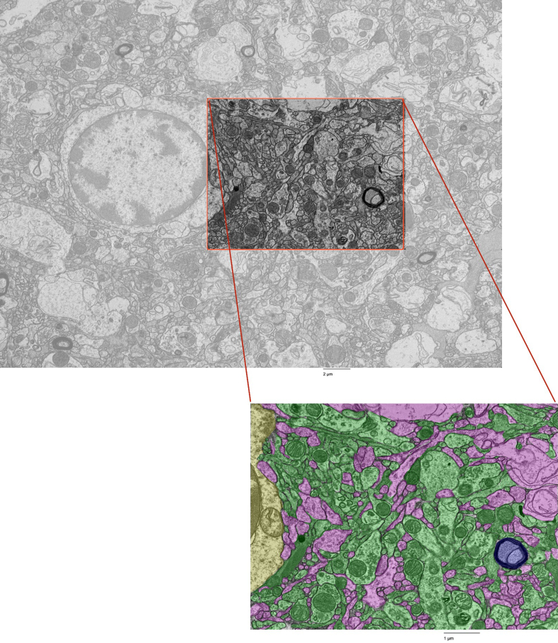
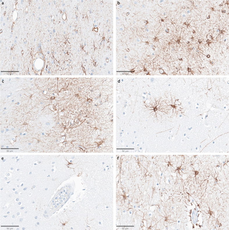
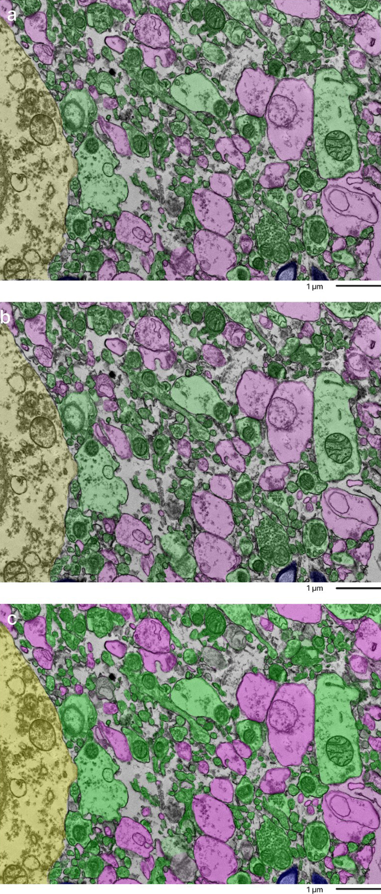
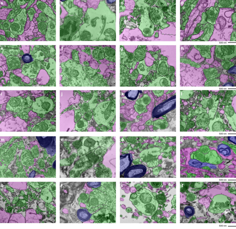
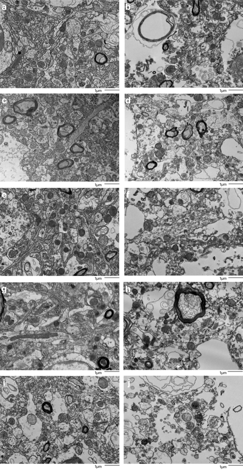
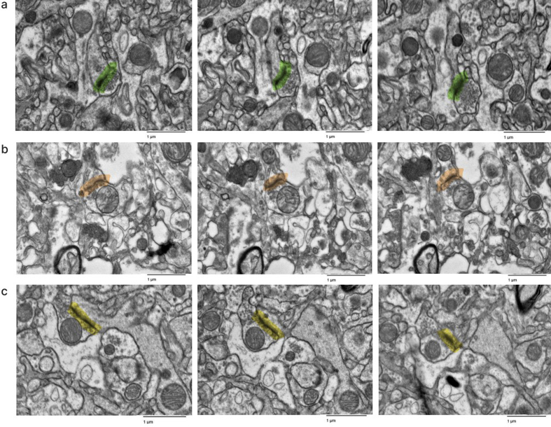
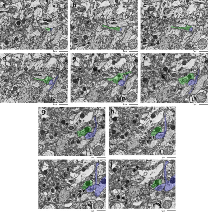
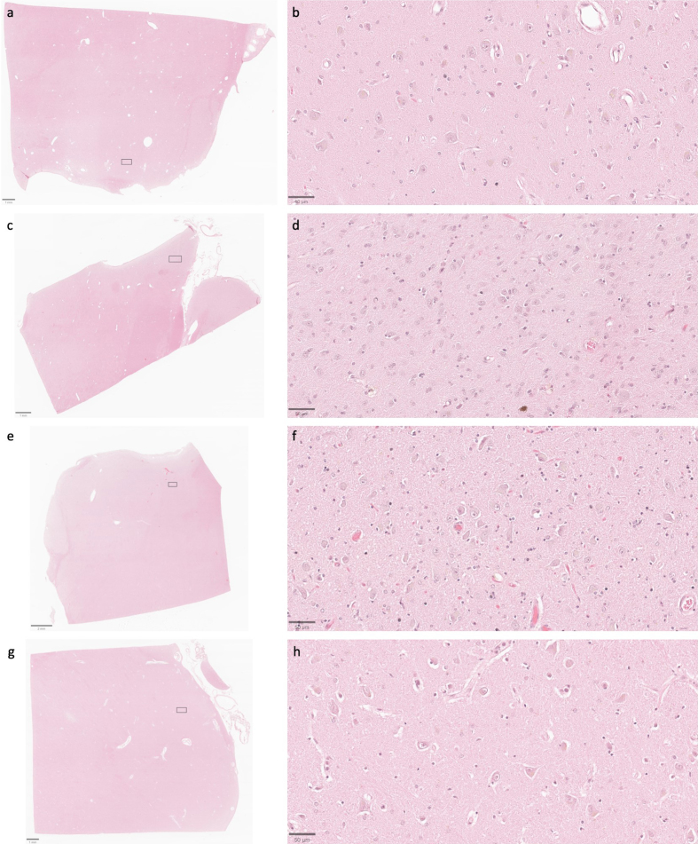
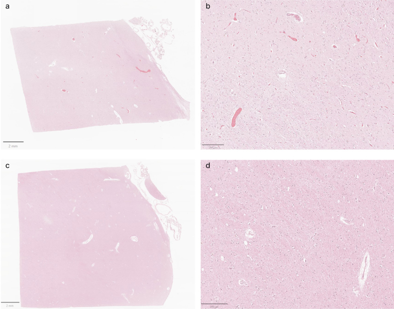
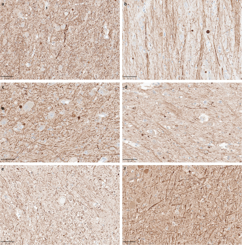
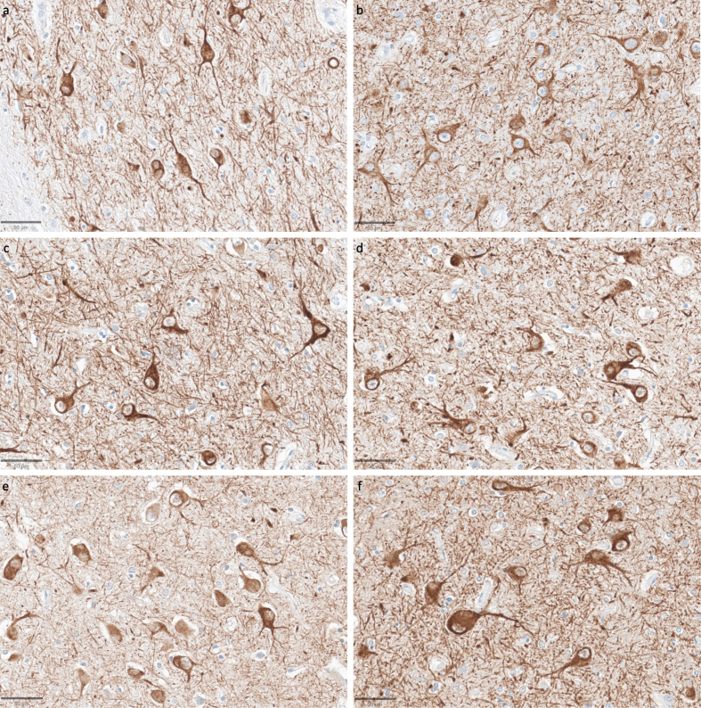
The ultrastructural analysis of postmortem brain tissue can provide important insights into cellular architecture and disease-related changes. For example, connectomics studies offer a powerful emerging approach for understanding neural circuit organization. However, electron microscopy (EM) data is difficult to interpret when the preservation quality is imperfect, which is common in brain banking and may render it unsuitable for certain research applications. One common issue is that EM images of postmortem brain tissue can have an expansion of regions that appear to be made up of extracellular space and / or degraded cellular material, which we call ambiguous interstitial zones. In this study, we report a method to assess whether EM images have ambiguous interstitial zone artifacts in a cohort of 10 postmortem brains with samples from each of the cortex and thalamus. Next, in matched samples from the contralateral hemisphere of the same brains, we evaluate the structural preservation quality of light microscopy images, including immunostaining for cytoskeletal proteins. Through this analysis, we show that on light microscopy, cell membrane morphology can be largely maintained, and neurite trajectory visualized over micrometer distances, even in specimens for which there are ambiguous interstitial zone artifacts on EM. Additionally, we demonstrate that synaptic structures can be successfully traced across serial EM sections in some postmortem samples, indicating the potential for connectivity studies in banked human brain tissue when appropriate preservation and visualization protocols are employed. Taken together, our analysis may assist in maximizing the usefulness of donated brain tissue by informing tissue selection and preparation protocols for various research goals.

摘要

对死后脑组织进行超微结构分析可以为细胞结构和疾病相关变化提供重要见解。例如,连接组学研究为理解神经回路组织提供了一种强大的新兴方法。然而,当保存质量不佳时,电子显微镜(EM)数据难以解读,这在脑库中很常见,可能使其不适用于某些研究应用。一个常见问题是,死后脑组织的EM图像可能会出现一些区域的扩张,这些区域似乎由细胞外空间和/或降解的细胞物质组成,我们称之为模糊间质区。在本研究中,我们报告了一种方法,用于评估10个死后大脑队列的EM图像是否存在模糊间质区伪影,这些大脑的样本取自皮质和丘脑。接下来,在同一大脑对侧半球的匹配样本中,我们评估光学显微镜图像的结构保存质量,包括细胞骨架蛋白的免疫染色。通过这项分析,我们表明,在光学显微镜下,即使在EM上存在模糊间质区伪影的标本中,细胞膜形态在很大程度上也能得以保持,神经突轨迹在微米距离上也能可视化。此外,我们证明,在一些死后样本中,可以成功地在连续的EM切片上追踪突触结构,这表明当采用适当的保存和可视化方案时,在保存的人类脑组织中进行连接性研究具有潜力。综上所述,我们的分析可以通过为各种研究目标提供组织选择和制备方案的信息,帮助最大限度地提高捐赠脑组织的有用性。

https://cdn.ncbi.nlm.nih.gov/pmc/blobs/026d/12189018/91b51637fe2e/freeneuropathol-06-13-6763-f11.jpg
https://cdn.ncbi.nlm.nih.gov/pmc/blobs/026d/12189018/2e2f01283e92/freeneuropathol-06-13-6763-f1.jpg
https://cdn.ncbi.nlm.nih.gov/pmc/blobs/026d/12189018/25491eb70133/freeneuropathol-06-13-6763-f2.jpg
https://cdn.ncbi.nlm.nih.gov/pmc/blobs/026d/12189018/1ec1fcef6065/freeneuropathol-06-13-6763-f3.jpg
https://cdn.ncbi.nlm.nih.gov/pmc/blobs/026d/12189018/845af5698594/freeneuropathol-06-13-6763-f4.jpg
https://cdn.ncbi.nlm.nih.gov/pmc/blobs/026d/12189018/9d1224a5f0a1/freeneuropathol-06-13-6763-f5.jpg
https://cdn.ncbi.nlm.nih.gov/pmc/blobs/026d/12189018/027afde8423b/freeneuropathol-06-13-6763-f6.jpg
https://cdn.ncbi.nlm.nih.gov/pmc/blobs/026d/12189018/766d5bcf2b28/freeneuropathol-06-13-6763-f7.jpg
https://cdn.ncbi.nlm.nih.gov/pmc/blobs/026d/12189018/60d58ddf444c/freeneuropathol-06-13-6763-f8.jpg
https://cdn.ncbi.nlm.nih.gov/pmc/blobs/026d/12189018/622204545b9b/freeneuropathol-06-13-6763-f9.jpg
https://cdn.ncbi.nlm.nih.gov/pmc/blobs/026d/12189018/61a2eb0462a2/freeneuropathol-06-13-6763-f10.jpg
https://cdn.ncbi.nlm.nih.gov/pmc/blobs/026d/12189018/91b51637fe2e/freeneuropathol-06-13-6763-f11.jpg

相似文献

1
Evaluating ultrastructural preservation quality in banked brain tissue.评估库存脑组织的超微结构保存质量。
Free Neuropathol. 2025 Jun 25;6:13. doi: 10.17879/freeneuropathology-2025-6763. eCollection 2025.
2
Evaluating ultrastructural preservation quality in banked brain tissue.评估库存脑组织的超微结构保存质量。
bioRxiv. 2025 May 14:2025.05.09.652503. doi: 10.1101/2025.05.09.652503.
3
Assessing the comparative effects of interventions in COPD: a tutorial on network meta-analysis for clinicians.评估慢性阻塞性肺疾病干预措施的比较效果:面向临床医生的网状Meta分析教程
Respir Res. 2024 Dec 21;25(1):438. doi: 10.1186/s12931-024-03056-x.
4
Comparison of cellulose, modified cellulose and synthetic membranes in the haemodialysis of patients with end-stage renal disease.纤维素、改性纤维素和合成膜在终末期肾病患者血液透析中的比较。
Cochrane Database Syst Rev. 2001(3):CD003234. doi: 10.1002/14651858.CD003234.
5
Systemic treatments for metastatic cutaneous melanoma.转移性皮肤黑色素瘤的全身治疗
Cochrane Database Syst Rev. 2018 Feb 6;2(2):CD011123. doi: 10.1002/14651858.CD011123.pub2.
6
Atypical antipsychotics for disruptive behaviour disorders in children and youths.用于治疗儿童和青少年破坏性行为障碍的非典型抗精神病药物。
Cochrane Database Syst Rev. 2017 Aug 9;8(8):CD008559. doi: 10.1002/14651858.CD008559.pub3.
7
Automated monitoring compared to standard care for the early detection of sepsis in critically ill patients.与标准护理相比,自动监测用于危重症患者脓毒症的早期检测
Cochrane Database Syst Rev. 2018 Jun 25;6(6):CD012404. doi: 10.1002/14651858.CD012404.pub2.
8
Direct composite resin fillings versus amalgam fillings for permanent posterior teeth.直接复合树脂充填与银汞合金充填用于永久性后牙。
Cochrane Database Syst Rev. 2021 Aug 13;8(8):CD005620. doi: 10.1002/14651858.CD005620.pub3.
9
A rapid and systematic review of the clinical effectiveness and cost-effectiveness of paclitaxel, docetaxel, gemcitabine and vinorelbine in non-small-cell lung cancer.对紫杉醇、多西他赛、吉西他滨和长春瑞滨在非小细胞肺癌中的临床疗效和成本效益进行的快速系统评价。
Health Technol Assess. 2001;5(32):1-195. doi: 10.3310/hta5320.
10
Cost-effectiveness of using prognostic information to select women with breast cancer for adjuvant systemic therapy.利用预后信息为乳腺癌患者选择辅助性全身治疗的成本效益
Health Technol Assess. 2006 Sep;10(34):iii-iv, ix-xi, 1-204. doi: 10.3310/hta10340.

本文引用的文献

1
Correlative light and electron microscopy for human brain and other biological models.用于人类大脑及其他生物模型的相关光电子显微镜技术。
Nat Protoc. 2025 Mar 31. doi: 10.1038/s41596-025-01153-9.
2
Ultrastructure of astrocytes using volume electron microscopy: A scoping review.使用体积电子显微镜观察星形胶质细胞的超微结构:一项范围综述。
J Physiol. 2025 Mar 3. doi: 10.1113/JP287455.
3
Volume electron microscopy reveals unique laminar synaptic characteristics in the human entorhinal cortex.体电子显微镜揭示了人类内嗅皮质独特的层状突触特征。
Elife. 2025 Jan 30;14:e96144. doi: 10.7554/eLife.96144.
4
A Drosophila computational brain model reveals sensorimotor processing.一个果蝇计算脑模型揭示了感觉运动处理过程。
Nature. 2024 Oct;634(8032):210-219. doi: 10.1038/s41586-024-07763-9. Epub 2024 Oct 2.
5
OsO as the Contrast-Generating Chemical Species of Osmium-Stained Biological Tissues in Electron Microscopy.OsO4 作为电子显微镜中锇染生物组织的对比生成化学物质。
Chembiochem. 2024 Oct 16;25(20):e202400311. doi: 10.1002/cbic.202400311. Epub 2024 Sep 12.
6
A petavoxel fragment of human cerebral cortex reconstructed at nanoscale resolution.重建的纳米分辨率人类大脑皮质 petavoxel 片段。
Science. 2024 May 10;384(6696):eadk4858. doi: 10.1126/science.adk4858.
7
Fluid preservation in brain banking: a review.脑库中的液体保存:综述
Free Neuropathol. 2024 Apr 23;5:10. doi: 10.17879/freeneuropathology-2024-5373. eCollection 2024 Jan.
8
Postmortem changes in brain cell structure: a review.脑细胞结构的死后变化:综述
Free Neuropathol. 2023 May 31;4:10. doi: 10.17879/freeneuropathology-2023-4790. eCollection 2023 Jan.
9
Pathological ultrastructural alterations of myelinated axons in normal appearing white matter in progressive multiple sclerosis.进行性多发性硬化症正常外观白质中髓鞘轴突的病理性超微结构改变。
Acta Neuropathol Commun. 2023 Jun 20;11(1):100. doi: 10.1186/s40478-023-01598-7.
10
Ex situ perfusion fixation for brain banking: a technical report.用于脑库的异位灌注固定:技术报告
Free Neuropathol. 2022 Sep 28;3:22. doi: 10.17879/freeneuropathology-2022-4368. eCollection 2022 Jan.