Li Fayuan, Gu Chengqi, Liang Chengpeng, Li Yang, Wang Shuo, Tang Qingqing, Jiang Huan, Linghu Shaorong, Dan Tingting, Shi Rong, Luo Xin, Liu Taixiang
Department of Ophthalmology, Affiliated Hospital of Zunyi Medical University, Zunyi, Guizhou Province, China.
Guizhou Provincial Branch of National Eye Disease Clinical Research Center, Zunyi, Guizhou Province, China.
Invest Ophthalmol Vis Sci. 2025 Jun 2;66(6):83. doi: 10.1167/iovs.66.6.83.
This study aims to construct a single-cell transcriptomic atlas of the developing rabbit sclera to elucidate fibroblast heterogeneity, differentiation trajectories, matrisome expression patterns, and intercellular communication, while revealing conserved molecular features of scleral cells through cross-species analysis.
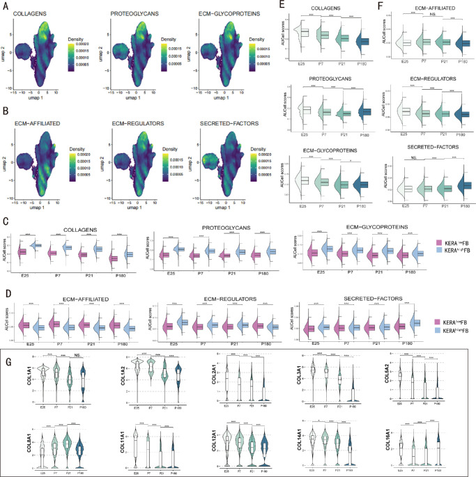
Single-cell RNA sequencing (scRNA-seq) was performed on scleral tissues from New Zealand rabbits at embryonic day 25 (E25) and postnatal days 7 (P7), 21 (P21), and 180 (P180). Libraries were prepared using the DNBelab C Series Kit and sequenced on the BGISEQ-2000 platform. Sequencing reads were aligned to the OryCun2.0 genome using STAR, and unique molecular identifier (UMI) count matrices were generated with PISA. Data preprocessing was conducted using Seurat. Fibroblast lineage differentiation was analyzed via VIA, intercellular communication via CellChat, matrisome expression patterns via AUCell, and cross-species analyses via CACIMAR and hdWGCNA.
We identified 7 major cell types and 15 subpopulations, with fibroblasts dominating the cellular landscape. Distinct fibroblast subtypes exhibited varied expression profiles and functions: KERAlow SPARCL1⁺ fibroblasts showed stem/progenitor-like features, while KERAhigh myocilin (MYOC)⁺ fibroblasts displayed senescence-associated phenotypes. Matrisome analysis revealed dynamic alterations in collagen and extracellular matrix (ECM)-related genes, and intercellular communication analysis highlighted complex signaling networks, particularly the MDK/PTN pathway. Cross-species comparisons demonstrated high conservation of fibroblasts between rabbit and human sclera, identifying four conserved co-expression modules.
This study presents the first single-cell atlas of rabbit scleral development, unveiling fibroblast heterogeneity, ECM remodeling mechanisms, and cross-species conserved features. These findings enhance our understanding of scleral biology and provide valuable insights for future research on ocular development and associated diseases, including myopia.
本研究旨在构建发育中的兔巩膜单细胞转录组图谱,以阐明成纤维细胞的异质性、分化轨迹、基质组表达模式和细胞间通讯,同时通过跨物种分析揭示巩膜细胞保守的分子特征。
对新西兰兔胚胎期第25天(E25)、出生后第7天(P7)、21天(P21)和180天(P180)的巩膜组织进行单细胞RNA测序(scRNA-seq)。使用DNBelab C系列试剂盒制备文库,并在BGISEQ-2000平台上进行测序。测序 reads 使用STAR 比对到OryCun2.0基因组,并使用PISA生成唯一分子标识符(UMI)计数矩阵。使用Seurat进行数据预处理。通过VIA分析成纤维细胞谱系分化,通过CellChat分析细胞间通讯,通过AUCell分析基质组表达模式,通过CACIMAR和hdWGCNA进行跨物种分析。
我们鉴定出7种主要细胞类型和15个亚群,其中成纤维细胞在细胞构成中占主导地位。不同的成纤维细胞亚型表现出不同的表达谱和功能:KERAlow SPARCL1⁺ 成纤维细胞表现出干细胞/祖细胞样特征,而KERAhigh 肌纤蛋白(MYOC)⁺ 成纤维细胞表现出衰老相关表型。基质组分析揭示了胶原蛋白和细胞外基质(ECM)相关基因的动态变化,细胞间通讯分析突出了复杂的信号网络,特别是MDK/PTN途径。跨物种比较表明兔和人巩膜中的成纤维细胞具有高度保守性,鉴定出四个保守的共表达模块。
本研究展示了首个兔巩膜发育的单细胞图谱,揭示了成纤维细胞的异质性、ECM重塑机制和跨物种保守特征。这些发现增进了我们对巩膜生物学的理解,并为未来关于眼发育及相关疾病(包括近视)的研究提供了有价值的见解。