• 文献检索
  • 文档翻译
  • 深度研究
  • 学术资讯
  • Suppr Zotero 插件Zotero 插件
  • 邀请有礼
  • 套餐&价格
  • 历史记录
应用&插件
Suppr Zotero 插件Zotero 插件浏览器插件Mac 客户端Windows 客户端微信小程序
定价
高级版会员购买积分包购买API积分包
服务
文献检索文档翻译深度研究API 文档MCP 服务
关于我们
关于 Suppr公司介绍联系我们用户协议隐私条款
关注我们

Suppr 超能文献

核心技术专利:CN118964589B侵权必究
粤ICP备2023148730 号-1Suppr @ 2026

文献检索

告别复杂PubMed语法,用中文像聊天一样搜索,搜遍4000万医学文献。AI智能推荐,让科研检索更轻松。

立即免费搜索

文件翻译

保留排版,准确专业,支持PDF/Word/PPT等文件格式,支持 12+语言互译。

免费翻译文档

深度研究

AI帮你快速写综述,25分钟生成高质量综述,智能提取关键信息,辅助科研写作。

立即免费体验

眼后部转录组分析完成了人类眼睛的细胞图谱。

Transcriptomic analysis of the ocular posterior segment completes a cell atlas of the human eye.

机构信息

Center for Brain Science, Harvard University, Cambridge, MA 02138.

F.M. Kirby Neurobiology Center, Boston Children's Hospital, Boston, MA 02115.

出版信息

Proc Natl Acad Sci U S A. 2023 Aug 22;120(34):e2306153120. doi: 10.1073/pnas.2306153120. Epub 2023 Aug 11.

DOI:10.1073/pnas.2306153120
PMID:37566633
原文链接:https://pmc.ncbi.nlm.nih.gov/articles/PMC10450437/
Abstract

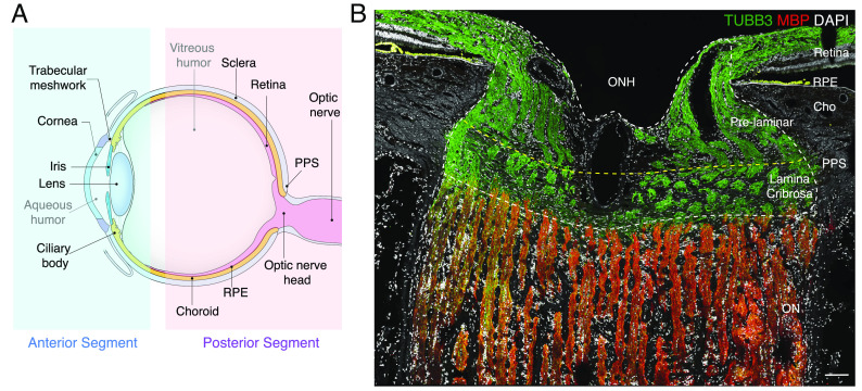
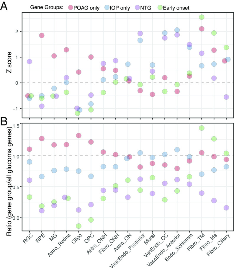
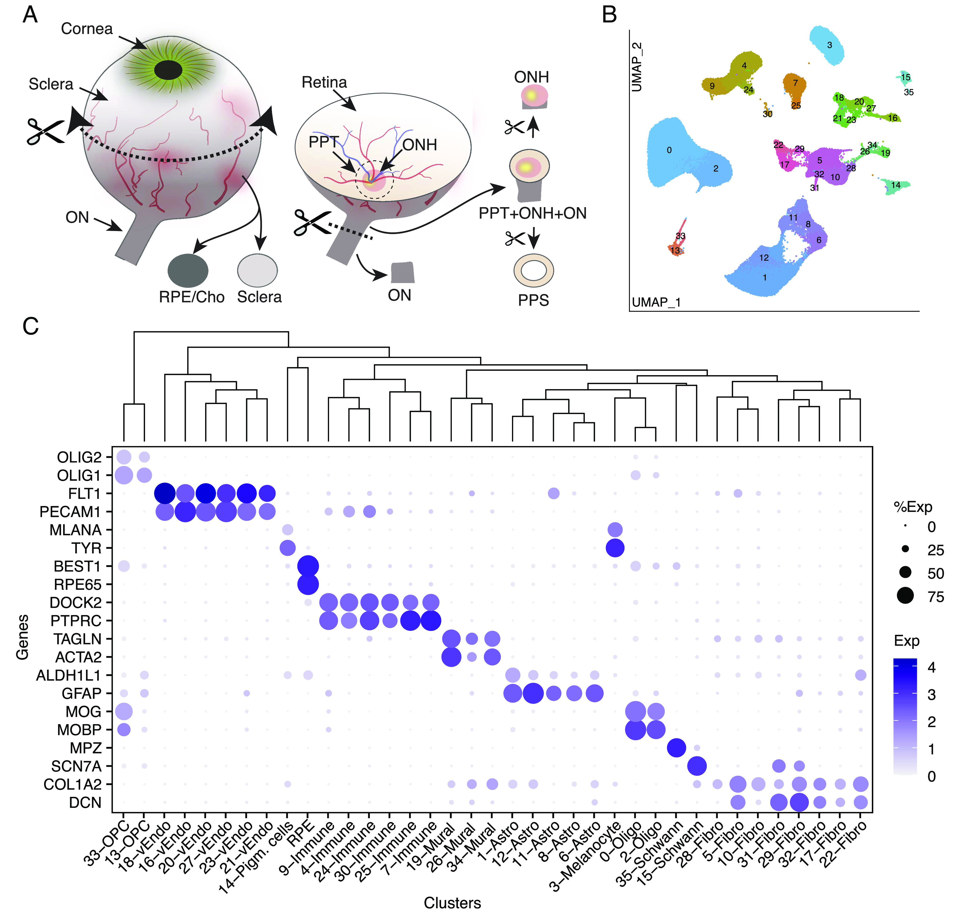
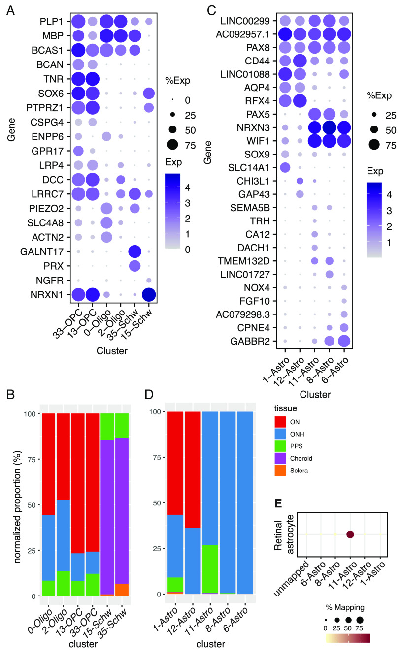
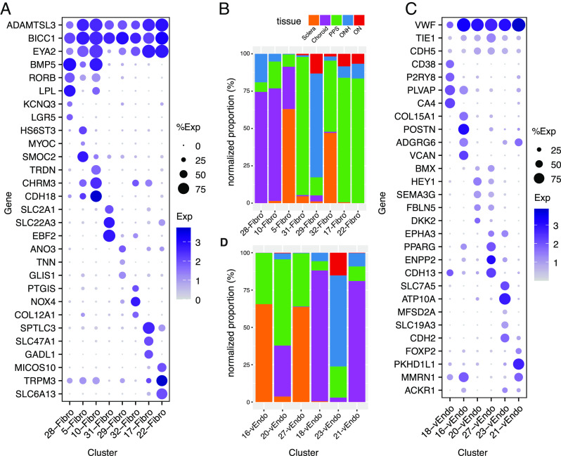
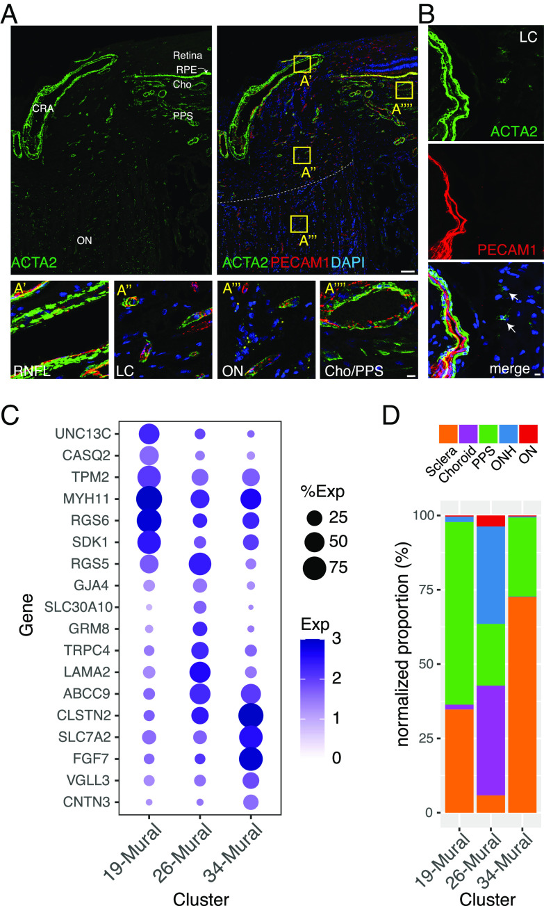
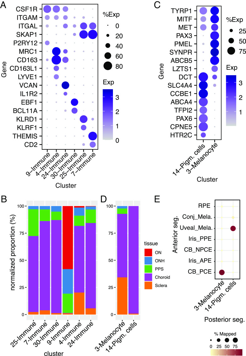
Although the visual system extends through the brain, most vision loss originates from defects in the eye. Its central element is the neural retina, which senses light, processes visual signals, and transmits them to the rest of the brain through the optic nerve (ON). Surrounding the retina are numerous other structures, conventionally divided into anterior and posterior segments. Here, we used high-throughput single-nucleus RNA sequencing (snRNA-seq) to classify and characterize cells in six extraretinal components of the posterior segment: ON, optic nerve head (ONH), peripheral sclera, peripapillary sclera (PPS), choroid, and retinal pigment epithelium (RPE). Defects in each of these tissues are associated with blinding diseases-for example, glaucoma (ONH and PPS), optic neuritis (ON), retinitis pigmentosa (RPE), and age-related macular degeneration (RPE and choroid). From ~151,000 single nuclei, we identified 37 transcriptomically distinct cell types, including multiple types of astrocytes, oligodendrocytes, fibroblasts, and vascular endothelial cells. Our analyses revealed a differential distribution of many cell types among distinct structures. Together with our previous analyses of the anterior segment and retina, the data presented here complete a "Version 1" cell atlas of the human eye. We used this atlas to map the expression of >180 genes associated with the risk of developing glaucoma, which is known to involve ocular tissues in both anterior and posterior segments as well as the neural retina. Similar methods can be used to investigate numerous additional ocular diseases, many of which are currently untreatable.

摘要

虽然视觉系统延伸到大脑,但大多数视力丧失都源于眼睛的缺陷。它的核心元素是神经视网膜,它感知光线,处理视觉信号,并通过视神经(ON)将它们传输到大脑的其他部分。视网膜周围有许多其他结构,传统上分为前段和后段。在这里,我们使用高通量单细胞 RNA 测序(snRNA-seq)对后段的六个眼外组织中的细胞进行分类和特征描述:视神经(ON)、视神经头(ONH)、周围巩膜、视盘周围巩膜(PPS)、脉络膜和视网膜色素上皮(RPE)。这些组织中的每一个组织的缺陷都与致盲性疾病有关,例如青光眼(ONH 和 PPS)、视神经炎(ON)、视网膜色素变性(RPE)和年龄相关性黄斑变性(RPE 和脉络膜)。从大约 151000 个单个核中,我们鉴定出 37 种转录上不同的细胞类型,包括多种类型的星形胶质细胞、少突胶质细胞、成纤维细胞和血管内皮细胞。我们的分析显示,许多细胞类型在不同结构中的分布存在差异。结合我们以前对前段和视网膜的分析,这里提供的数据完成了人类眼睛的“版本 1”细胞图谱。我们使用该图谱来绘制与青光眼风险相关的>180 个基因的表达图谱,已知青光眼涉及前段和后段的眼部组织以及神经视网膜。类似的方法可用于研究许多其他眼部疾病,其中许多目前尚无治疗方法。

https://cdn.ncbi.nlm.nih.gov/pmc/blobs/ab26/10450437/46117193b362/pnas.2306153120fig07.jpg
https://cdn.ncbi.nlm.nih.gov/pmc/blobs/ab26/10450437/645011f3e158/pnas.2306153120fig01.jpg
https://cdn.ncbi.nlm.nih.gov/pmc/blobs/ab26/10450437/00f3fea64781/pnas.2306153120fig02.jpg
https://cdn.ncbi.nlm.nih.gov/pmc/blobs/ab26/10450437/f2ac1e2b35b6/pnas.2306153120fig03.jpg
https://cdn.ncbi.nlm.nih.gov/pmc/blobs/ab26/10450437/415ba54061f5/pnas.2306153120fig04.jpg
https://cdn.ncbi.nlm.nih.gov/pmc/blobs/ab26/10450437/a32de831e6c1/pnas.2306153120fig05.jpg
https://cdn.ncbi.nlm.nih.gov/pmc/blobs/ab26/10450437/5d049ef08d63/pnas.2306153120fig06.jpg
https://cdn.ncbi.nlm.nih.gov/pmc/blobs/ab26/10450437/46117193b362/pnas.2306153120fig07.jpg

相似文献

1
Transcriptomic analysis of the ocular posterior segment completes a cell atlas of the human eye.眼后部转录组分析完成了人类眼睛的细胞图谱。
Proc Natl Acad Sci U S A. 2023 Aug 22;120(34):e2306153120. doi: 10.1073/pnas.2306153120. Epub 2023 Aug 11.
2
Transcriptomic Analysis of the Ocular Posterior Segment Completes a Cell Atlas of the Human Eye.眼后段的转录组分析完成了人类眼睛的细胞图谱。
bioRxiv. 2023 Apr 27:2023.04.26.538447. doi: 10.1101/2023.04.26.538447.
3
Transcriptomic analysis across nasal, temporal, and macular regions of human neural retina and RPE/choroid by RNA-Seq.通过RNA测序对人类神经视网膜以及视网膜色素上皮/脉络膜的鼻侧、颞侧和黄斑区域进行转录组分析。
Exp Eye Res. 2014 Dec;129:93-106. doi: 10.1016/j.exer.2014.11.001. Epub 2014 Nov 5.
4
3-D histomorphometry of the normal and early glaucomatous monkey optic nerve head: lamina cribrosa and peripapillary scleral position and thickness.正常及早期青光眼猴视神经乳头的三维组织形态计量学:筛板及视乳头周围巩膜的位置和厚度
Invest Ophthalmol Vis Sci. 2007 Oct;48(10):4597-607. doi: 10.1167/iovs.07-0349.
5
Comparative quantitative study of astrocytes and capillary distribution in optic nerve laminar regions.视神经层区星形胶质细胞与毛细血管分布的比较定量研究。
Exp Eye Res. 2014 Apr;121:11-22. doi: 10.1016/j.exer.2014.02.008. Epub 2014 Feb 19.
6
Biomechanics of the optic nerve head and sclera in canine glaucoma: A brief review.犬青光眼视神经乳头和巩膜的生物力学:简要综述。
Vet Ophthalmol. 2021 Jul;24(4):316-325. doi: 10.1111/vop.12923. Epub 2021 Aug 17.
7
Regional Deformation of the Optic Nerve Head and Peripapillary Sclera During IOP Elevation.眼压升高时视乳头和视盘周围巩膜的区域性变形。
Invest Ophthalmol Vis Sci. 2018 Jul 2;59(8):3779-3788. doi: 10.1167/iovs.18-24462.
8
Differing Associations between Optic Nerve Head Strains and Visual Field Loss in Patients with Normal- and High-Tension Glaucoma.正常眼压性青光眼和高眼压性青光眼患者视神经乳头应变与视野缺损之间的不同关联。
Ophthalmology. 2023 Jan;130(1):99-110. doi: 10.1016/j.ophtha.2022.08.007. Epub 2022 Aug 11.
9
IOP-induced regional displacements in the optic nerve head and correlation with peripapillary sclera thickness.IOP 诱导的视神经头区域位移与视盘周围巩膜厚度的相关性。
Exp Eye Res. 2020 Nov;200:108202. doi: 10.1016/j.exer.2020.108202. Epub 2020 Aug 27.
10
Enhanced depth imaging optical coherence tomography of deep optic nerve complex structures in glaucoma.青光眼深层视神经复合体结构的增强深度成像光相干断层扫描。
Ophthalmology. 2012 Jan;119(1):3-9. doi: 10.1016/j.ophtha.2011.07.012. Epub 2011 Oct 5.

引用本文的文献

1
Analysis of 2-dimensional regional differences in the peripapillary scleral fibroblast cytoskeleton of normotensive and hypertensive mouse eyes.正常血压和高血压小鼠眼视乳头周围巩膜成纤维细胞细胞骨架的二维区域差异分析。
Sci Rep. 2025 Jul 1;15(1):21207. doi: 10.1038/s41598-025-08251-4.
2
Single-Cell RNA Sequencing of Rabbit Sclera at Different Developmental Stages: Unveiling Scleral Cells Atlas and the Heterogeneity of Fibroblasts.不同发育阶段兔巩膜的单细胞RNA测序:揭示巩膜细胞图谱和成纤维细胞的异质性
Invest Ophthalmol Vis Sci. 2025 Jun 2;66(6):83. doi: 10.1167/iovs.66.6.83.
3
Distinct Transcriptomic Profiles of Cultured Anterior and Posterior Populations of Human Infant Scleral Fibroblasts: Including Dopamine Receptors.

本文引用的文献

1
Evolution of neuronal cell classes and types in the vertebrate retina.脊椎动物视网膜中神经元细胞类型和类别的演化。
Nature. 2023 Dec;624(7991):415-424. doi: 10.1038/s41586-023-06638-9. Epub 2023 Dec 13.
2
Dynamic human retinal pigment epithelium (RPE) and choroid architecture based on single-cell transcriptomic landscape analysis.基于单细胞转录组景观分析的动态人类视网膜色素上皮(RPE)和脉络膜结构
Genes Dis. 2022 Dec 15;10(6):2540-2556. doi: 10.1016/j.gendis.2022.11.007. eCollection 2023 Nov.
3
Transcriptomic and Chromatin Accessibility Analysis of the Human Macular and Peripheral Retinal Pigment Epithelium at the Single-Cell Level.
人类婴儿巩膜成纤维细胞前体和后体培养物的不同转录组特征:包括多巴胺受体
Invest Ophthalmol Vis Sci. 2025 May 1;66(5):29. doi: 10.1167/iovs.66.5.29.
4
Paired DNA and RNA sequencing uncovers common and rare genetic variants regulating gene expression in the human retina.配对DNA和RNA测序揭示了调控人类视网膜基因表达的常见和罕见遗传变异。
medRxiv. 2025 Apr 26:2025.04.25.25326445. doi: 10.1101/2025.04.25.25326445.
5
Optineurin-facilitated axonal mitochondria delivery promotes neuroprotection and axon regeneration.视紫质促进轴突线粒体运输,从而促进神经保护和轴突再生。
Nat Commun. 2025 Feb 20;16(1):1789. doi: 10.1038/s41467-025-57135-8.
6
Multi-omic spatial effects on high-resolution AI-derived retinal thickness.多组学空间效应对高分辨率人工智能衍生的视网膜厚度的影响。
Nat Commun. 2025 Feb 4;16(1):1317. doi: 10.1038/s41467-024-55635-7.
7
Cysteinyl leukotriene receptor 1 modulates retinal immune cells, vascularity and proteolytic activity in aged mice.半胱氨酰白三烯受体1调节老年小鼠的视网膜免疫细胞、血管生成和蛋白水解活性。
Aging (Albany NY). 2025 Jan 31;17(2):308-328. doi: 10.18632/aging.206193.
8
Multiparametric grading of glaucoma severity by histopathology can enable post-mortem substratification of disease state.通过组织病理学对青光眼严重程度进行多参数分级可以实现疾病状态的死后分层。
Acta Neuropathol Commun. 2025 Jan 6;13(1):3. doi: 10.1186/s40478-024-01880-2.
9
Genetic Insights into Therapeutic Targets for Neuromyelitis Optica Spectrum Disorders: A Mendelian Randomization Study.视神经脊髓炎谱系障碍治疗靶点的遗传学见解:一项孟德尔随机化研究
Mol Neurobiol. 2025 May;62(5):5518-5530. doi: 10.1007/s12035-024-04612-8. Epub 2024 Nov 20.
10
Base-excision repair pathway shapes 5-methylcytosine deamination signatures in pan-cancer genomes.碱基切除修复通路塑造泛癌症基因组中 5-甲基胞嘧啶脱氨酶的特征。
Nat Commun. 2024 Nov 14;15(1):9864. doi: 10.1038/s41467-024-54223-z.
单细胞水平下人眼黄斑和周边视网膜色素上皮的转录组学和染色质可及性分析。
Am J Pathol. 2023 Nov;193(11):1750-1761. doi: 10.1016/j.ajpath.2023.01.012. Epub 2023 Feb 11.
4
Single-cell RNA sequencing reveals transcriptional changes of human choroidal and retinal pigment epithelium cells during fetal development, in healthy adult and intermediate age-related macular degeneration.单细胞 RNA 测序揭示了人类脉络膜和视网膜色素上皮细胞在胎儿发育、健康成人和年龄相关性黄斑变性中期的转录变化。
Hum Mol Genet. 2023 May 5;32(10):1698-1710. doi: 10.1093/hmg/ddad007.
5
Thrombospondin 1 missense alleles induce extracellular matrix protein aggregation and TM dysfunction in congenital glaucoma.血栓反应蛋白 1 错义等位基因导致先天性青光眼细胞外基质蛋白聚集和 TM 功能障碍。
J Clin Invest. 2022 Dec 1;132(23):e156967. doi: 10.1172/JCI156967.
6
Insights from DOCK2 in cell function and pathophysiology.DOCK2在细胞功能和病理生理学中的见解。
Front Mol Biosci. 2022 Sep 29;9:997659. doi: 10.3389/fmolb.2022.997659. eCollection 2022.
7
Remodeling of the Lamina Cribrosa: Mechanisms and Potential Therapeutic Approaches for Glaucoma.筛板重塑:青光眼的潜在治疗方法及机制。
Int J Mol Sci. 2022 Jul 22;23(15):8068. doi: 10.3390/ijms23158068.
8
Cell atlas of the human ocular anterior segment: Tissue-specific and shared cell types.人类眼前节细胞图谱:组织特异性和共享细胞类型。
Proc Natl Acad Sci U S A. 2022 Jul 19;119(29):e2200914119. doi: 10.1073/pnas.2200914119. Epub 2022 Jul 15.
9
Regional and age-related diversity of human mature oligodendrocytes.人类成熟少突胶质细胞的区域和年龄相关性多样性。
Glia. 2022 Oct;70(10):1938-1949. doi: 10.1002/glia.24230. Epub 2022 Jun 23.
10
Single Cell Transcriptomic Analysis Reveals Organ Specific Pericyte Markers and Identities.单细胞转录组分析揭示器官特异性周细胞标志物和特性。
Front Cardiovasc Med. 2022 Jun 1;9:876591. doi: 10.3389/fcvm.2022.876591. eCollection 2022.