Mohammadi Hedayatullah, Güneş İrfan Botan, Arı Şeyhmus
Department of Ophthalmology, Medicalpark Kocaeli Hospital, 41050, Kocaeli, Turkey.
Medicalpark Kocaeli Hospital, Department of Ophthalmology, Kocaeli Health and Technology University, Kocaeli, 41245, Turkey.
BMC Ophthalmol. 2025 Jul 1;25(1):361. doi: 10.1186/s12886-025-04184-8.
Various studies have shown decreased retinal blood flow in patients with active Graves' orbitopathy (GO). We investigated whether retinal blood flow returned to normal levels after intravenous glucocorticoid treatment in active GO patients.
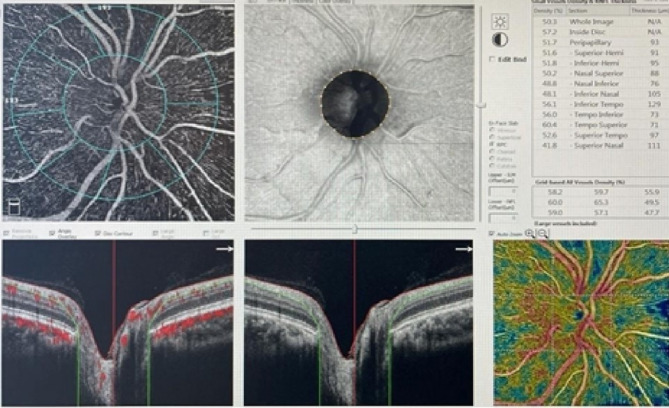
Thirty-two eyes of 32 patients with moderate or severe active GO and 32 eyes of 32 healthy participants were included. The blood flow in the superficial plexus (SP), deep retinal plexus (DP), external retinal (ER) capillary plexus and choriocapillaris was measured via OCT-Angiography before treatment with methylprednisolone and one week after the completion of treatment. In addition, central macular thickness, intraocular pressure, and best corrected visual acuity, were compared with those of the control group.
Retinal blood flow (RBF) in the macula at SP, DP, ER and choriocapillaris levels was 48.38 ± 2.04, 54.17 ± 3.11, 0.42 ± 0.14 and 2176.45 ± 147.53 in the control group and 45.91 ± 4.56, 51.55 ± 3.84, 0.35 ± 0.34 and 2030.15 ± 132.15 in the active GO group, respectively. The RBF was significantly lower in the active GO group than in the control group at all angiographic levels (p < 0.001). Although blood flow increased after treatment, it remained lower than that in the control group (p < 0.001). The mean CMT was significantly greater in active GO patients before treatment than in the controls and after treatment (p < 0.001). No serious systemic or ocular side effects were observed in any of the patients.
After pulse glucocorticoid treatment, retinal blood flow increases but remains low compared with that in the healthy control group.
多项研究表明,活动性格雷夫斯眼眶病(GO)患者的视网膜血流减少。我们调查了活动性GO患者静脉注射糖皮质激素治疗后视网膜血流是否恢复到正常水平。
纳入32例中度或重度活动性GO患者的32只眼和32名健康参与者的32只眼。在用甲泼尼龙治疗前和治疗完成后一周,通过光学相干断层扫描血管造影术测量浅表丛(SP)、视网膜深层丛(DP)、视网膜外(ER)毛细血管丛和脉络膜毛细血管的血流。此外,将中心黄斑厚度、眼压和最佳矫正视力与对照组进行比较。
对照组SP、DP、ER和脉络膜毛细血管水平黄斑区的视网膜血流(RBF)分别为48.38±2.04、54.17±3.11、0.42±0.14和2176.45±147.53,活动性GO组分别为45.91±4.56、51.55±3.84、0.35±0.34和2030.15±132.15。在所有血管造影水平上,活动性GO组的RBF均显著低于对照组(p<0.001)。虽然治疗后血流增加,但仍低于对照组(p<0.001)。活动性GO患者治疗前的平均中心黄斑厚度显著大于对照组和治疗后(p<0.001)。所有患者均未观察到严重的全身或眼部副作用。
脉冲糖皮质激素治疗后,视网膜血流增加,但与健康对照组相比仍较低。