Wang Cheng, Sun Jinchun, Donakonda Rohini, Beger Richard, Latham Leah E, Wu Leihong, Liu Shuliang, Hanig Joseph P, Liu Fang
Division of Neurotoxicology, National Center for Toxicological Research/FDA, Jefferson, AR, United States.
Division of Systems Biology, National Center for Toxicological Research/U.S. Food and Drug Administration (FDA), Jefferson, AR, United States.
Exp Biol Med (Maywood). 2025 Jun 25;250:10607. doi: 10.3389/ebm.2025.10607. eCollection 2025.
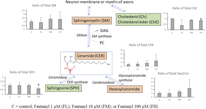
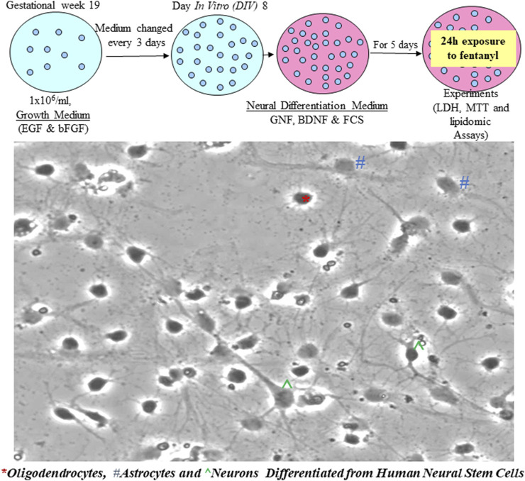
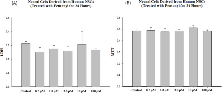
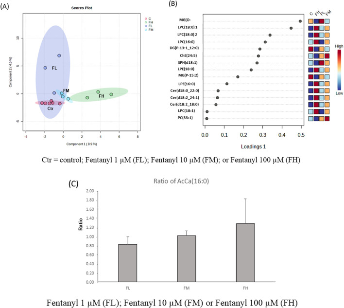
Fentanyl is a potent and short-acting opioid that is often given to pediatric patients during surgery to relieve pain and as an adjunct to anesthesia. Its effects on the developing brain are yet to be determined. In the present study, commercially available human neural stem cells (NSCs) were used to model the effects of fentanyl on the developing human brain. We determined the dose dependent effects and temporal relationships between fentanyl exposures and NSC health, viability, and differentiation. Markers of mitochondrial health [3-(4,5-dimethylthiazol-2-yl)-2,5-diphenyltetra-zolium bromide (MTT)] and cell death/damage [lactate dehydrogenase (LDH)] were monitored to determine the dose response effects of fentanyl on NSC viability. In addition, lipidomics analysis was conducted to investigate lipid profile changes in differentiated neural cells treated with fentanyl. Fentanyl did not cause a significant increase in LDH release, nor MTT reduction after 24-h exposure at concentrations of 0.5, 1.0, 3.0, 10, or 100 μM, for both NSCs and differentiated neural cells. Lipidomics data showed the top 15 most variable important in projection (VIP) lipid species (the higher the VIP scores, the bigger changes in treated groups vs. controls), including lysophosphatidylcholines (LPCs), lysophosphatidylethanolamines (LPEs), ceramides (CER), cholesterol esters (ChEs) and sphingosine (SPH). The lipidomic data indicate that LPC (16:0), LPC (16:1), LPC (18:1), CER (d18:0_22:0), CER (d18:2_18:0), CER(d18:2_24:1) were significantly increased, and only ChE (24:5) and SPH (d18:1) were significantly decreased in the highest dose group versus control. These data indicated that fentanyl exposure (24-h) did not induce detectable cell death. However, a lipidomic analysis indicated that fentanyl may affect immature neural cell functions through modifying lipid composition and lipid metabolism. These data indicated that despite the absence of clear neurodegeneration, fentanyl may still have a negative impact on the developing brain.
芬太尼是一种强效短效阿片类药物,常用于小儿手术患者以缓解疼痛并作为麻醉辅助剂。其对发育中大脑的影响尚待确定。在本研究中,使用市售的人神经干细胞(NSCs)来模拟芬太尼对发育中人类大脑的影响。我们确定了芬太尼暴露与神经干细胞健康、活力及分化之间的剂量依赖性效应和时间关系。监测线粒体健康标志物[3-(4,5-二甲基噻唑-2-基)-2,5-二苯基四氮唑溴盐(MTT)]和细胞死亡/损伤标志物[乳酸脱氢酶(LDH)],以确定芬太尼对神经干细胞活力的剂量反应效应。此外,进行脂质组学分析以研究用芬太尼处理的分化神经细胞中的脂质谱变化。对于神经干细胞和分化神经细胞,在0.5、1.0、3.0、10或100μM浓度下暴露24小时后,芬太尼未导致LDH释放显著增加,也未导致MTT还原。脂质组学数据显示了投影中最重要的15种变化最大的脂质种类(VIP脂质种类,VIP分数越高,处理组与对照组的变化越大),包括溶血磷脂酰胆碱(LPCs)、溶血磷脂酰乙醇胺(LPEs)、神经酰胺(CER)、胆固醇酯(ChEs)和鞘氨醇(SPH)。脂质组学数据表明,在最高剂量组与对照组相比,LPC(16:0)、LPC(16:1)、LPC(18:1)、CER(d18:0_22:0)、CER(d18:2_18:0)、CER(d18:2_24:1)显著增加,而仅ChE(24:5)和SPH(d18:1)显著降低。这些数据表明,芬太尼暴露(24小时)未诱导可检测到的细胞死亡。然而,脂质组学分析表明,芬太尼可能通过改变脂质组成和脂质代谢来影响未成熟神经细胞功能。这些数据表明,尽管没有明显的神经退行性变,芬太尼仍可能对发育中的大脑产生负面影响。