Stoldt Stefan, Maass Frederike, Weber Michael, Dennerlein Sven, Ilgen Peter, Gärtner Jutta, Canfes Aysenur, Schweighofer Sarah V, Jans Daniel C, Rehling Peter, Jakobs Stefan
Department of NanoBiophotonics, Max Planck Institute for Multidisciplinary Sciences, RG Mitochondrial Structure and Dynamics, Göttingen, Germany.
Clinic of Neurology, University Medical Center Göttingen, Göttingen, Germany.
Nat Commun. 2025 Jul 10;16(1):6391. doi: 10.1038/s41467-025-61577-5.
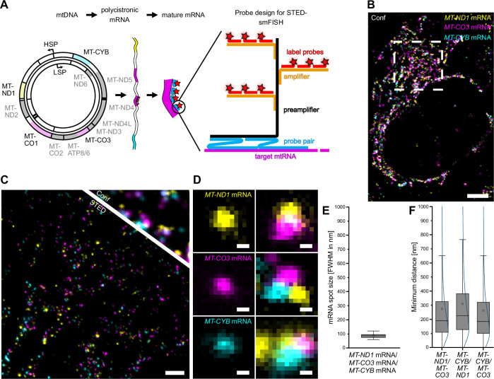
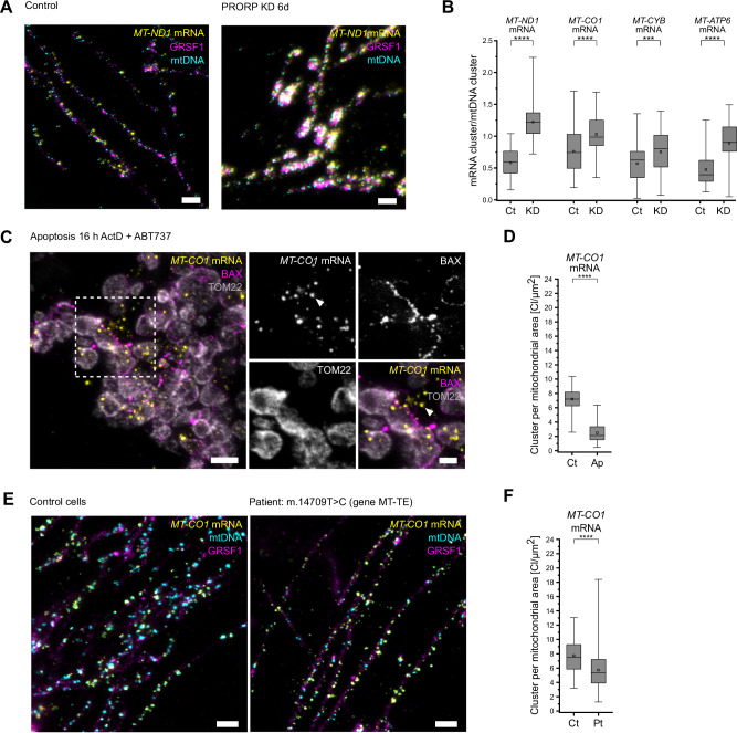
Mitochondria contain their own DNA (mtDNA) and a dedicated gene expression machinery. As the mitochondrial dimensions are close to the diffraction limit of classical light microscopy, the spatial distribution of mitochondrial proteins and in particular of mitochondrial mRNAs remains underexplored. Here, we establish single-molecule fluorescence in situ hybridization (smFISH) combined with STED and MINFLUX super-resolution microscopy (nanoscopy) to visualize individual mitochondrial mRNA molecules and associated proteins. STED nanoscopy reveals the spatial relationships between distinct mRNA species and proteins such as the RNA granule marker GRSF1, demonstrating adaptive changes in mRNA distribution and quantity in challenged mammalian cells and patient-derived cell lines. Notably, STED-smFISH shows the release of mRNAs during apoptosis, while MINFLUX reveals the folding of the mRNAs into variable shapes, as well as their spatial proximity to mitochondrial ribosomes. These protocols are transferable to various cell types and open new avenues for understanding mitochondrial gene regulation in health and disease.
线粒体含有自己的DNA(mtDNA)和一套专门的基因表达机制。由于线粒体的尺寸接近传统光学显微镜的衍射极限,线粒体蛋白质尤其是线粒体mRNA的空间分布仍未得到充分研究。在这里,我们建立了单分子荧光原位杂交(smFISH),并结合受激发射损耗(STED)和最小荧光涨落成像(MINFLUX)超分辨率显微镜(纳米显微镜)来可视化单个线粒体mRNA分子和相关蛋白质。STED纳米显微镜揭示了不同mRNA种类与诸如RNA颗粒标记物GRSF1等蛋白质之间的空间关系,证明了在受到挑战的哺乳动物细胞和患者来源的细胞系中mRNA分布和数量的适应性变化。值得注意的是,STED-smFISH显示了凋亡过程中mRNA的释放,而MINFLUX揭示了mRNA折叠成可变形状以及它们与线粒体核糖体的空间接近度。这些方法可转移到各种细胞类型,并为理解健康和疾病状态下的线粒体基因调控开辟了新途径。