Mura Rebecca, Pochepnia Svitlana, Kifjak Daria, Khenkina Natallia, Prosch Helmut
Department of Medicine and Surgery (diMeC), University of Parma, Parma 43125, Italy.
Christian Doppler Laboratory for Machine Learning Driven Precision Imaging, Department of Biomedical Imaging and Image-guided Therapy, Medical University of Vienna, Vienna 1090, Austria.
BJR Open. 2025 May 8;7(1):tzaf009. doi: 10.1093/bjro/tzaf009. eCollection 2025 Jan.
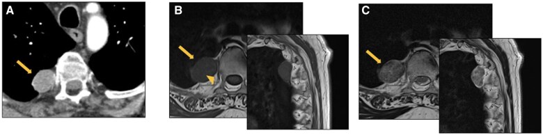
Mediastinal masses represent a heterogeneous group of entities characterized by a variety of histopathological and radiological features. Imaging plays a pivotal role in the detection and interpretation of mediastinal abnormalities. CT remains the modality of choice due to its high spatial and temporal resolution and its ability to assess tissue composition, including the detection of fluid, fat, and calcifications. MRI represents a complementary tool in specific scenarios, such as differentiating complicated cysts from solid lesions or identifying intracellular fat content, as seen in thymic hyperplasia. The differential diagnosis of mediastinal masses relies primarily on the location of the mass and tissue composition, integrated with clinical characteristics of the patient. This review discusses the most common mediastinal masses in adults, providing a practical approach to their differentiation mainly based on the predominant density pattern and location.
纵隔肿块是一组异质性病变,具有多种组织病理学和放射学特征。影像学在纵隔异常的检测和解读中起着关键作用。由于CT具有高空间和时间分辨率,以及评估组织成分的能力,包括检测液体、脂肪和钙化,因此它仍然是首选的检查方式。MRI在特定情况下是一种辅助工具,例如区分复杂囊肿与实性病变或识别细胞内脂肪含量,如胸腺增生所见。纵隔肿块的鉴别诊断主要依赖于肿块的位置和组织成分,并结合患者的临床特征。本综述讨论了成人最常见的纵隔肿块,主要基于主要密度模式和位置提供了一种实用的鉴别方法。