Lang Blerta, Boden Karl, Stanzel Boris V, Prothmann Ulrich, Szurman Peter
Eye Clinic Sulzbach, Knappschaft Kliniken Saar GmbH, An der Klinik 10, Sulzbach, D-66280, Germany.
Klaus Heimann Eye Research Institute, An der Klinik 10, Sulzbach/Saar, D-66280, Germany.
J Ophthalmic Inflamm Infect. 2025 Jul 22;15(1):57. doi: 10.1186/s12348-025-00519-0.
Acute zonal occult outer retinopathy (AZOOR) is a rare inflammatory disease of the outer retina, often presented with subtle early findings. A specific subtype, termed Multizonal Outer Retinopathy and Retinal Pigment Epitheliopathy (MORR), is characterized by distinct progression pattern (Ramtohul et al. Retina 43:1890–1903, 2023) in multiple zones of the outer retina and retinal pigment epithelium. This case report aims to illustrate the chronologically divergent presentation, a phase-shifting disease progression and the complex clinical course of MORR, and to discuss the diagnostic challenges posed by its phase‑shifted timeline as well as potential therapeutic options.
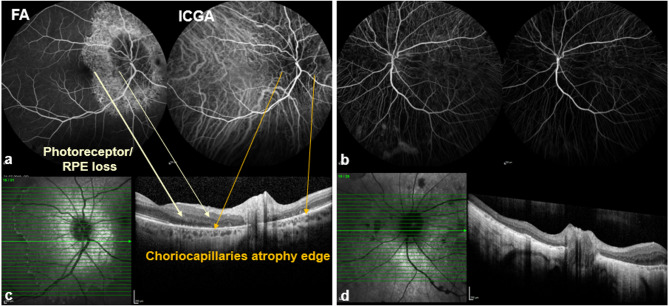
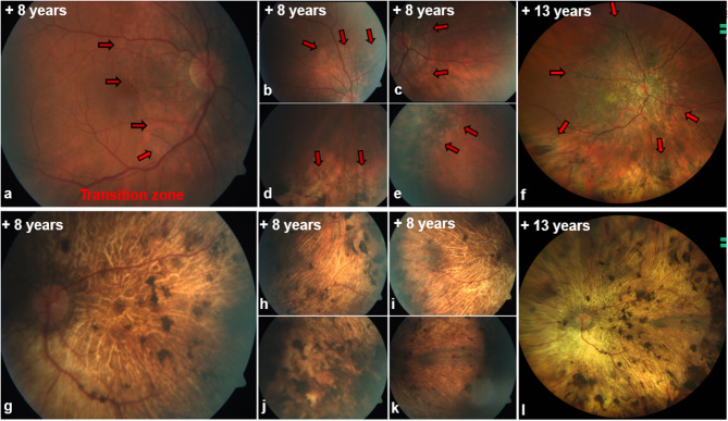
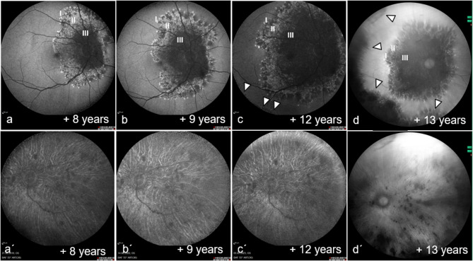
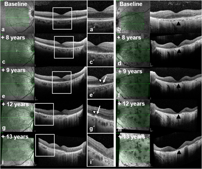
A case of a 52-year-old female patient with initially unilateral, inactive posterior uveitis was retrospectively analyzed. Over thirteen years, progressive functional impairment developed in the fellow eye. Findings were assessed using multimodal imaging (Optical Coherence Tomography [OCT], Fundus Autofluorescence [FAF], Fluorescein and Indocyanine Green Angiography [FFA/ICGA]) and electrophysiological examinations (multifocal ERG, full-field ERG, EOG, VEP). Additional rheumatologic and neurologic assessments were conducted, and an infectious workup was performed.
The patient demonstrated chronologically divergent bilateral involvement with extensive damage to the retinal pigment epithelium (RPE) and photoreceptors. Fundus autofluorescence revealed a tri- to multizonal pattern in the better eye, while the fellow eye already exhibited diffuse atrophic areas devoid of any autofluorescence. Electrophysiological, the better eye showed selectively prolonged latencies on multifocal electroretinography (multifocal ERG) but preserved amplitudes, whereas the more severely affected eye displayed substantial functional loss. Despite various therapeutic interventions, including high-dose corticosteroids and immunosuppressive agents, progressive visual impairment ensued, driven by increasing macular involvement.
This case highlights the marked heterogeneity and diagnostic complexities of Multizonal Outer Retinopathy and Retinal Pigment Epitheliopathy (MORR), a newly recognized progressive variant of Acute Zonal Occult Outer Retinopathy (AZOOR). The disease course can present with chronologically divergent manifestations in both eyes. While initial stages may exhibit only subtle funduscopic changes, structural and functional deficits can progress rapidly, episodically and in a phase-shifted manner. Multimodal imaging is essential to delineate the disease trajectory and to distinguish MORR from diseases affecting outer retina. Currently, no definitive treatment is available; although immunomodulatory therapies may stabilize the condition in certain cases, their efficacy remains inconsistent. Consequently, early low-vision management and close interdisciplinary collaboration are of particular importance.
急性区域性隐匿性外层视网膜病变(AZOOR)是一种罕见的外层视网膜炎症性疾病,早期表现往往较为隐匿。一种特定的亚型,称为多区域外层视网膜病变和视网膜色素上皮病变(MORR),其特征是外层视网膜和视网膜色素上皮的多个区域呈现出独特的进展模式(Ramtohul等人,《视网膜》43:1890 - 1903,2023)。本病例报告旨在阐述MORR按时间顺序出现的不同表现、疾病进展的相移以及复杂的临床过程,并讨论其相移时间线带来的诊断挑战以及潜在的治疗选择。
对一名52岁女性患者的病例进行回顾性分析,该患者最初表现为单侧、非活动性后葡萄膜炎。在13年的时间里,对侧眼出现了进行性的功能损害。使用多模态成像(光学相干断层扫描[OCT]、眼底自发荧光[FAF]、荧光素和吲哚菁绿血管造影[FFA/ICGA])和电生理检查(多焦视网膜电图、全视野视网膜电图、眼电图、视觉诱发电位)对检查结果进行评估。还进行了额外的风湿病学和神经学评估,并进行了感染性检查。
该患者表现出按时间顺序出现的双侧不同程度受累,视网膜色素上皮(RPE)和光感受器广泛受损。眼底自发荧光显示较好眼呈现三区域至多区域模式,而对侧眼已经出现弥漫性萎缩区域,无任何自发荧光。电生理方面,较好眼在多焦视网膜电图(多焦ERG)上显示选择性潜伏期延长但振幅保留,而受影响更严重的眼则表现出明显的功能丧失。尽管进行了各种治疗干预,包括高剂量皮质类固醇和免疫抑制剂,但由于黄斑受累增加,视力仍逐渐下降。
本病例突出了多区域外层视网膜病变和视网膜色素上皮病变(MORR)的显著异质性和诊断复杂性,MORR是急性区域性隐匿性外层视网膜病变(AZOOR)新认识的一种进行性变体。疾病过程在双眼可能表现出按时间顺序出现的不同表现。虽然初始阶段可能仅表现为细微的眼底改变,但结构和功能缺陷可能迅速、间歇性且以相移的方式进展。多模态成像对于描绘疾病轨迹以及将MORR与影响外层视网膜的疾病区分开来至关重要。目前尚无确切的治疗方法;尽管免疫调节疗法在某些情况下可能使病情稳定,但其疗效仍不一致。因此,早期低视力管理和密切的跨学科合作尤为重要。