文献检索文档翻译深度研究
Suppr Zotero 插件Zotero 插件
邀请有礼套餐&价格历史记录

新学期,新优惠

限时优惠:9月1日-9月22日

30天高级会员仅需29元

1天体验卡首发特惠仅需5.99元

了解详情
不再提醒
插件&应用
Suppr Zotero 插件Zotero 插件浏览器插件Mac 客户端Windows 客户端微信小程序
高级版
套餐订阅购买积分包
AI 工具
文献检索文档翻译深度研究
关于我们
关于 Suppr公司介绍联系我们用户协议隐私条款
关注我们

Suppr 超能文献

核心技术专利:CN118964589B侵权必究
粤ICP备2023148730 号-1Suppr @ 2025

脊髓损伤后血浆细胞外囊泡谱随时间和严重程度的变化

Temporal and Severity-Dependent Alterations in Plasma Extracellular Vesicle Profiles Following Spinal Cord Injury.

作者信息

Cooper Jamie, Airey Scott Tait, Patino Eric, Andriot Theo, Ghosh Mousumi, Pearse Damien D

机构信息

The Miami Project to Cure Paralysis, Department of Neurological Surgery, University of Miami Miller School of Medicine, Miami, FL 33136, USA.

The Neuroscience Program, University of Miami Miller School of Medicine, Miami, FL 33136, USA.

出版信息

Cells. 2025 Jul 11;14(14):1065. doi: 10.3390/cells14141065.


DOI:10.3390/cells14141065
PMID:40710318
原文链接:https://pmc.ncbi.nlm.nih.gov/articles/PMC12294027/
Abstract

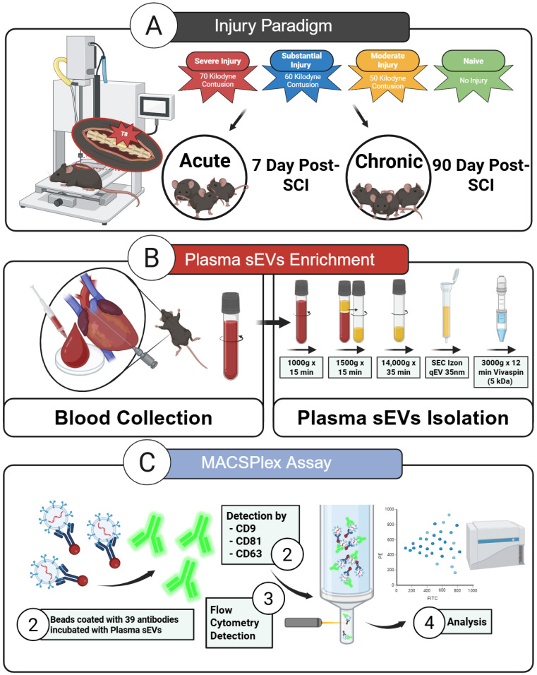
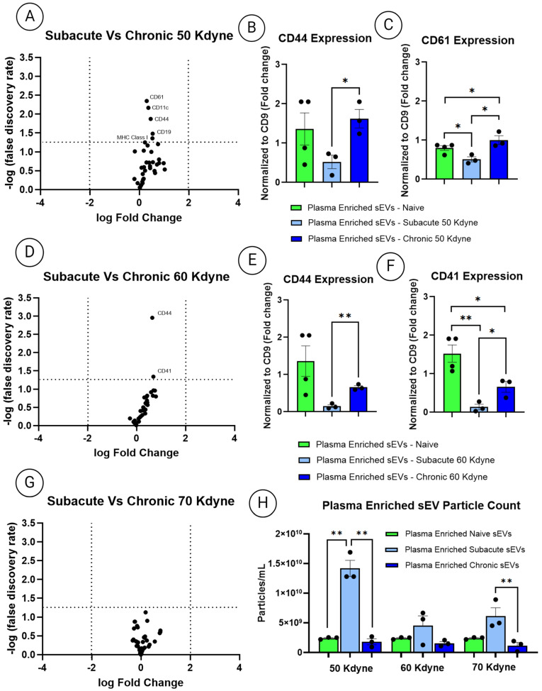
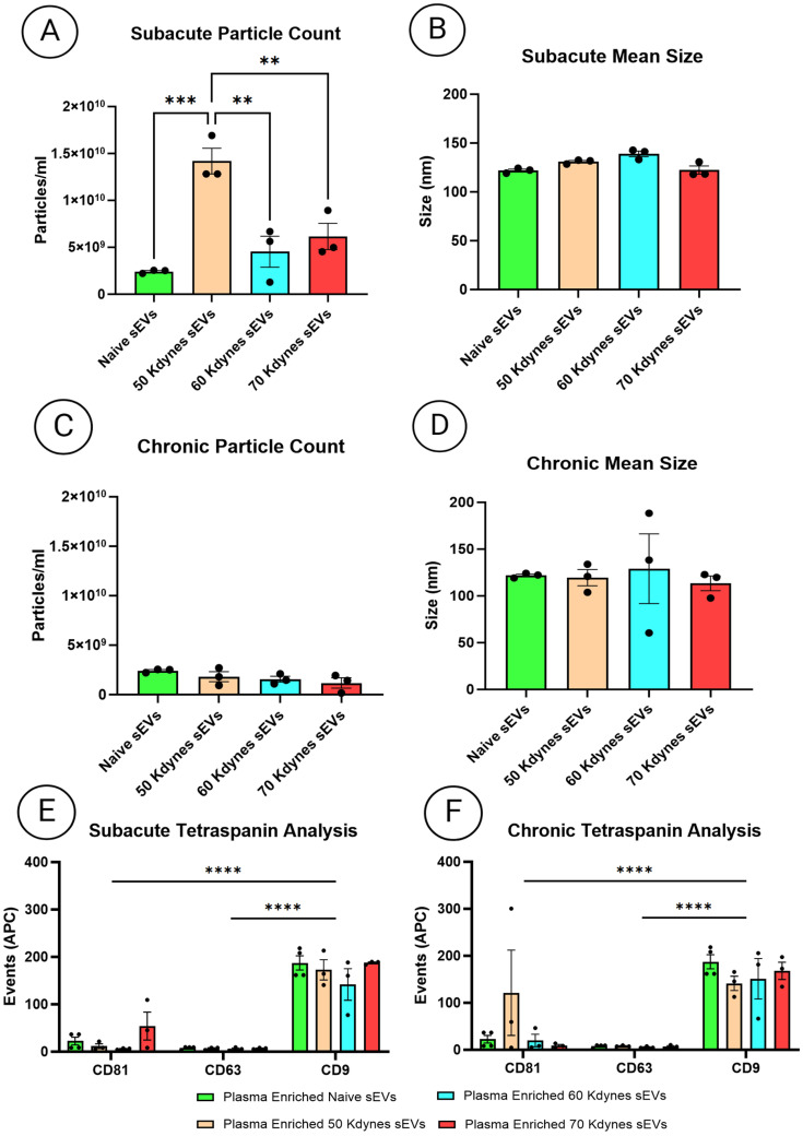
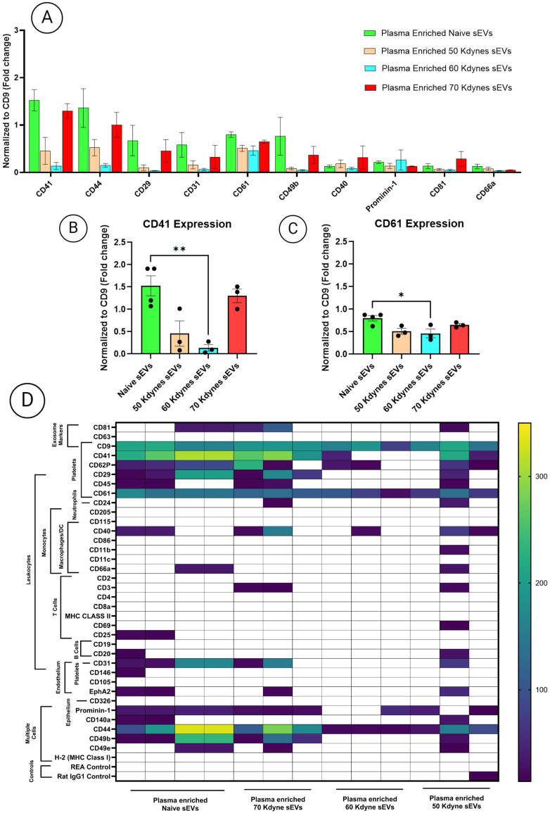
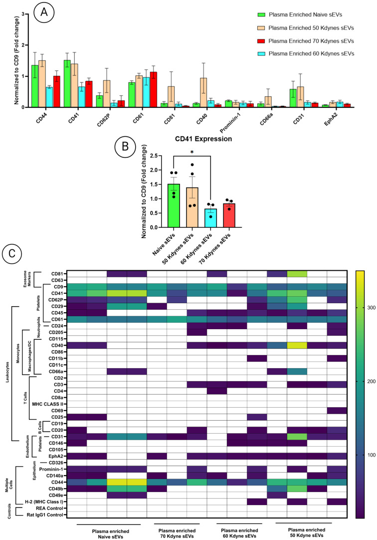
Spinal cord injury (SCI) triggers both local and systemic pathological responses that evolve over time and differ with injury severity. Small extracellular vesicles (sEVs), known mediators of intercellular communication, may serve as biomarkers reflecting these complex dynamics. In this study, we investigated whether SCI severity modulates the composition and abundance of circulating plasma-derived sEVs across subacute and chronic phases. Using a graded thoracic contusion model in mice, plasma was collected at defined timepoints post-injury. sEVs were isolated via size-exclusion chromatography and characterized using nanoparticle tracking analysis (NTA), transmission electron microscopy (TEM), and MACSPlex surface marker profiling. We observed an SCI-dependent increase in sEVs during the subacute (7 days) phase, most notably in moderate injuries (50 kdyne), with overall vesicle counts lower chronically (3 months). CD9 emerged as the predominant tetraspanin sEV marker, while CD63 and CD81 were generally present at low levels across all injury severities and timepoints. Surface sEV analysis revealed dynamic regulation of CD41, CD44, and CD61 in the CD9 sEV subset, suggesting persistent systemic signaling activity. These markers, traditionally associated with platelet function, may also reflect immune or reparative responses following SCI. Our findings highlight the evolving nature of sEV profiles after SCI and support their potential as non-invasive biomarkers for monitoring injury progression.

摘要

脊髓损伤(SCI)会引发局部和全身的病理反应,这些反应会随时间演变且因损伤严重程度而异。小细胞外囊泡(sEVs)作为细胞间通讯的已知介质,可能充当反映这些复杂动态变化的生物标志物。在本研究中,我们调查了SCI严重程度是否会在亚急性期和慢性期调节循环血浆来源的sEVs的组成和丰度。使用小鼠分级胸椎挫伤模型,在损伤后的特定时间点收集血浆。通过尺寸排阻色谱法分离sEVs,并使用纳米颗粒跟踪分析(NTA)、透射电子显微镜(TEM)和MACSPlex表面标志物分析对其进行表征。我们观察到在亚急性期(7天)sEVs出现了依赖于SCI的增加,最明显的是在中度损伤(50达因)中,而慢性期(3个月)的总体囊泡计数较低。CD9成为主要的四跨膜蛋白sEV标志物,而CD63和CD81在所有损伤严重程度和时间点的水平通常较低。表面sEV分析揭示了CD9 sEV亚群中CD41、CD44和CD61的动态调节,表明存在持续的全身信号活动。这些传统上与血小板功能相关的标志物,也可能反映SCI后的免疫或修复反应。我们的研究结果突出了SCI后sEV谱的演变性质,并支持它们作为监测损伤进展的非侵入性生物标志物的潜力。

https://cdn.ncbi.nlm.nih.gov/pmc/blobs/198e/12294027/5df52ac7c7f5/cells-14-01065-g007.jpg
https://cdn.ncbi.nlm.nih.gov/pmc/blobs/198e/12294027/58364d48e852/cells-14-01065-g001.jpg
https://cdn.ncbi.nlm.nih.gov/pmc/blobs/198e/12294027/68220dca782a/cells-14-01065-g002.jpg
https://cdn.ncbi.nlm.nih.gov/pmc/blobs/198e/12294027/5da3793e822e/cells-14-01065-g003.jpg
https://cdn.ncbi.nlm.nih.gov/pmc/blobs/198e/12294027/2231c72c75ed/cells-14-01065-g004.jpg
https://cdn.ncbi.nlm.nih.gov/pmc/blobs/198e/12294027/e261dba08b3f/cells-14-01065-g005.jpg
https://cdn.ncbi.nlm.nih.gov/pmc/blobs/198e/12294027/b3b8f200a7ca/cells-14-01065-g006.jpg
https://cdn.ncbi.nlm.nih.gov/pmc/blobs/198e/12294027/5df52ac7c7f5/cells-14-01065-g007.jpg

相似文献

[1]
Temporal and Severity-Dependent Alterations in Plasma Extracellular Vesicle Profiles Following Spinal Cord Injury.

Cells. 2025-7-11

[2]
Single vesicle analysis reveals the release of tetraspanin positive extracellular vesicles into circulation with high intensity intermittent exercise.

J Physiol. 2023-11

[3]
Engineered small extracellular vesicles for targeted delivery of perlecan to stabilise the blood-spinal cord barrier after spinal cord injury.

Clin Transl Med. 2025-6

[4]
Human Infrapatellar Fat Pad Mesenchymal Stem Cell-derived Extracellular Vesicles Purified by Anion Exchange Chromatography Suppress Osteoarthritis Progression in a Mouse Model.

Clin Orthop Relat Res. 2024-7-1

[5]
Diverse Populations of Extracellular Vesicles with Opposite Functions during Herpes Simplex Virus 1 Infection.

J Virol. 2021-2-24

[6]
Temporal changes in the protein cargo of extracellular vesicles and resultant immune reprogramming after severe burn injury in humans and mice.

Front Immunol. 2025-6-4

[7]
Multi-omics uncovers immune-modulatory molecules in plasma contributing to resistance exercise-ameliorated locomotor disability after incomplete spinal cord injury.

Genome Med. 2025-2-5

[8]
Systemic pharmacological treatments for chronic plaque psoriasis: a network meta-analysis.

Cochrane Database Syst Rev. 2021-4-19

[9]
Establishing Indicative Neurofilament Gradients Based on Severity of Spinal Cord Injury.

World Neurosurg. 2025-3

[10]
[Effect of removing microglia from spinal cord on nerve repair after spinal cord injury in mice].

Zhongguo Xiu Fu Chong Jian Wai Ke Za Zhi. 2025-6-15

本文引用的文献

[1]
Rab27a-mediated extracellular vesicle release drives astrocytic CSPG secretion and glial scarring in spinal cord injury.

Biomater Adv. 2025-11

[2]
Control of Physical and Biochemical Parameters Influencing Exogeneous Cargo Protein Association to Extracellular Vesicles Using Lipid Anchors Enables High Loading and Effective Intracellular Delivery.

J Extracell Biol. 2025-5-14

[3]
Extracellular vesicles epitopes as potential biomarker candidates in patients with traumatic spinal cord injury.

Front Immunol. 2024-11-27

[4]
The Yin and Yang of Microglia-Derived Extracellular Vesicles in CNS Injury and Diseases.

Cells. 2024-11-6

[5]
Reduced circulating CD63 extracellular vesicle levels associate with atherosclerosis in hypercholesterolaemic mice and humans.

Cardiovasc Diabetol. 2024-10-17

[6]
Comparison of inferior vena cava puncture under continuous cardiac perfusion with cardiac puncture in blood acquisition of the laboratory mouse.

Lab Anim. 2025-4

[7]
The role of neuroinflammation in neurodegenerative diseases: current understanding and future therapeutic targets.

Front Aging Neurosci. 2024-4-12

[8]
Minimal information for studies of extracellular vesicles (MISEV2023): From basic to advanced approaches.

J Extracell Vesicles. 2024-2

[9]
Inhibition of CD44 suppresses the formation of fibrotic scar after spinal cord injury via the JAK2/STAT3 signaling pathway.

iScience. 2024-1-16

[10]
Schwann Cell-Derived Exosomal Vesicles: A Promising Therapy for the Injured Spinal Cord.

Int J Mol Sci. 2023-12-10

文献AI研究员

20分钟写一篇综述,助力文献阅读效率提升50倍

立即体验

用中文搜PubMed

大模型驱动的PubMed中文搜索引擎

马上搜索

推荐工具

医学文档翻译智能文献检索