Cingoz Eda, Cingoz Mehmet, Comert Rana Gunoz, Dursun Memduh
Radiology Department, Bagcilar Training and Research Hospital, Istanbul, Turkey.
Radiology Department, Basaksehir Cam and Sakura City Hospital, Istanbul, Turkey.
Medicine (Baltimore). 2025 Jul 25;104(30):e43586. doi: 10.1097/MD.0000000000043586.
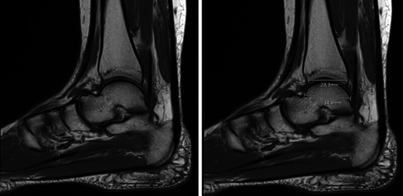
Lateral osteochondral lesions of the talus (OLT) are a notable cause of chronic ankle pain, particularly in cases without a history of trauma. However, their anatomical risk factors remain understudied compared to medial OLTs. This study aimed to identify anatomical factors associated with the development of nontraumatic lateral OLT using magnetic resonance imaging (MRI)-based morphometric measurements. MRI scans from 62 ankles were examined in this retrospective study, comprising 31 patients with lateral OLT and 31 healthy controls matched by age, sex, and side. The following parameters were measured on the MR images: the anterior opening angle of the talus (AOT), the angle between the tibial axis and medial malleolus (TMM), the angle of the tibial plafond to the malleolus, the angle between the anterior and posterior tibiofibular ligaments (ATFL-PTFL angle), the length of the distal tibial articular surface (TAS), the length of the trochlea tali arc (TAL), the ratio of TAS to TAL (TAS/TAL), and the depth of the incisura fibularis (IncDep). AOT, IncDep, ATFL-PTFL angle, TMM, TAL, and TAS/TAL demonstrated significant differences between the 2 groups (P < .05). The threshold values were as follows: 13.5° (area under the curve [AUC] 0.807) for AOT, 80.5° (AUC 0.767) for ATFL-PTFL angle, 16.5° (AUC 0.920) for TMM, and 0.80 (AUC 0.704) for TAS/TAL. Multivariate logistic regression analysis indicated an odds ratio (OR) = 17.805 for AOT ≥ 13.5°, OR = 19.887 for ATFL-PTFL angle > 80.5°, OR = 27.576 for TMM > 1.5° and OR = 4.680 for TAS/TAL ≤ 0.80. These findings suggest that specific anatomical parameters identifiable on MRI, particularly increased AOT, TMM, and ATFL-PTFL angle, are significantly associated with the development of nontraumatic lateral OLT. These parameters may serve as useful imaging biomarkers for clinical risk assessment.
距骨外侧骨软骨损伤(OLT)是慢性踝关节疼痛的一个显著原因,尤其是在没有创伤史的病例中。然而,与内侧OLT相比,其解剖学危险因素仍未得到充分研究。本研究旨在通过基于磁共振成像(MRI)的形态测量来确定与非创伤性外侧OLT发生相关的解剖学因素。在这项回顾性研究中,检查了62个踝关节的MRI扫描图像,包括31例外侧OLT患者和31名年龄、性别及患侧相匹配的健康对照者。在MR图像上测量了以下参数:距骨前开口角(AOT)、胫骨轴线与内踝之间的角度(TMM)、胫骨平台与内踝的角度、胫腓前韧带与胫腓后韧带之间的角度(ATFL-PTFL角)、胫骨远端关节面长度(TAS)、距骨滑车弧长度(TAL)、TAS与TAL的比值(TAS/TAL)以及腓骨切迹深度(IncDep)。AOT、IncDep、ATFL-PTFL角、TMM、TAL和TAS/TAL在两组之间存在显著差异(P < 0.05)。阈值如下:AOT为13.5°(曲线下面积[AUC] 0.807),ATFL-PTFL角为80.5°(AUC 0.767),TMM为16.5°(AUC 0.920),TAS/TAL为0.80(AUC 0.704)。多因素逻辑回归分析表明,AOT≥13.5°时比值比(OR) = 17.805,ATFL-PTFL角>80.5°时OR = 19.887,TMM>16.5°时OR = 27.576,TAS/TAL≤0.80时OR = 4.680。这些发现表明,MRI上可识别的特定解剖学参数,特别是增大的AOT、TMM以及ATFL-PTFL角,与非创伤性外侧OLT的发生显著相关。这些参数可作为临床风险评估的有用影像生物标志物。