Cao Mingtai, Liu Xinyi, Yang Airu, Xu Yuan, Zhang Qian, Cao Yuntai
Department of Radiology, Affiliated Hospital of Qinghai University, Xining, China.
Department of Radiology, The Second Hospital & Clinical Medical School, Lanzhou University, Lanzhou, China.
Gland Surg. 2025 Jul 31;14(7):1195-1212. doi: 10.21037/gs-2025-83. Epub 2025 Jul 28.
Breast cancer remains the predominant contributor to global cancer-related morbidity and mortality in women. Luminal subtypes, accounting for approximately 70% of cases, demonstrate favorable prognoses through endocrine-targeted therapeutic regimens owing to hormone receptor positivity. Conversely, non-luminal breast cancer variants, including human epidermal growth factor receptor 2 (HER2)-enriched and triple-negative subtypes, exhibit aggressive biological characteristics, intrinsic endocrine therapy resistance, and require molecularly guided therapeutic strategies such as HER2-directed biologicals, platinum-based cytotoxic regimens, or radiation therapy. This study aims to evaluate whether preoperative multiparametric magnetic resonance imaging (MRI)-based intratumoral and peritumoral radiomics can effectively discriminate between luminal and non-luminal breast cancer subtypes.
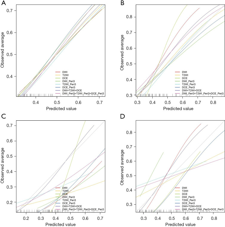
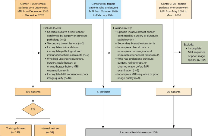
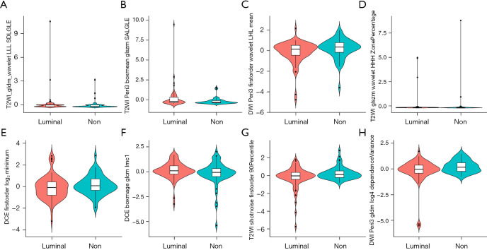
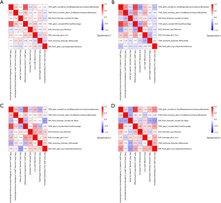
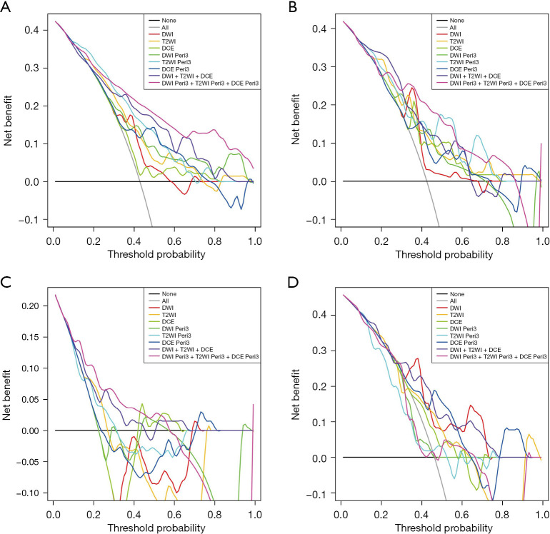
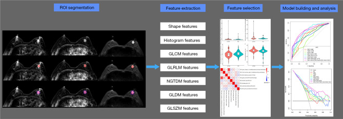
This retrospective study analyzed 305 female breast cancer patients. Center 1 (Affiliated Hospital of Qinghai University) was randomly split into a training set (n=140) and an internal test set (n=59) in a 7:3 ratio, while Center 2 (Second Hospital of Lanzhou University) (n=67) and Center 3 (The Cancer Imaging Archive I-SPY1 trial) (n=39) served as external test sets 1 and 2, respectively. Tumor subtypes were classified as luminal or non-luminal based on estrogen receptor (ER) and progesterone receptor (PR) status. Two radiologists performed manual tumor segmentation using 3D Slicer on multiparametric MRI sequences: dynamic contrast enhancement (DCE; phases 3 or 4), fat-suppressed T2-weighted imaging (T2WI), and diffusion-weighted imaging (DWI). Peritumoral regions were defined by a 3 mm expansion from the tumor volume of interest (VOI). For each sequence (intratumoral and peritumoral), 2,252 radiomics features were extracted using PyRadiomics. After Z-score normalization, features were selected through univariate analysis, correlation analysis, and simulated annealing. Eight radiomics models were constructed using random forest (RF), including intratumoral-only, combined intratumoral-peritumoral (3 mm), and multisequence fusion models. Performance was assessed using area under the curve (AUC), calibration curves, and decision curve analysis (DCA).
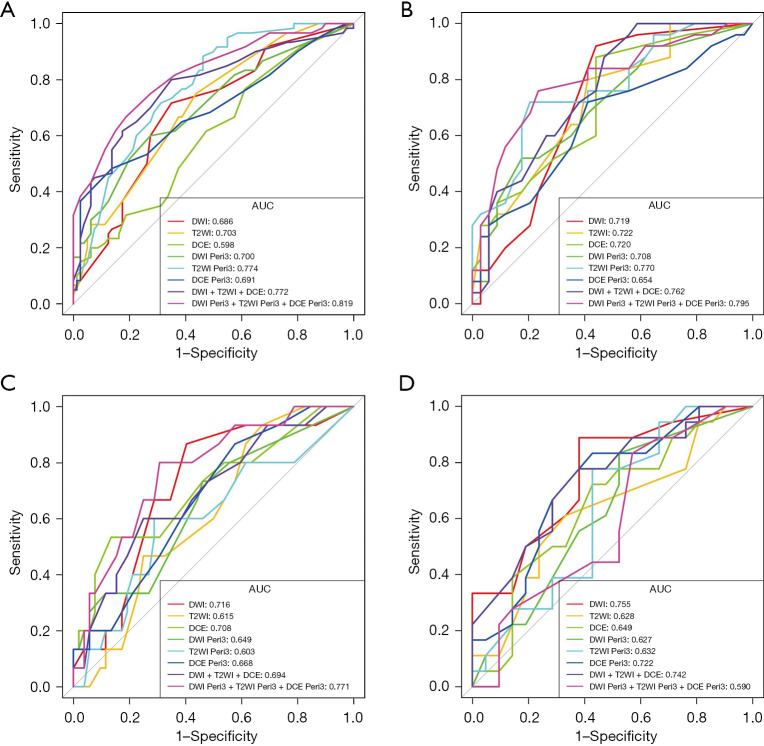
After feature selection, eight optimal radiomics features were used for model development. The combined DWI_Peri3 + T2WI_Peri3 + DCE_Peri3 RF model demonstrated superior performance, with AUCs of 0.819 [95% confidence interval (CI): 0.748-0.889], 0.795 (95% CI: 0.676-0.915), and 0.771 (95% CI: 0.640-0.902) in training, internal validation, and external validation set 1, respectively. Among single-parameter models, T2WI_Peri3 RF showed the best classification performance (AUC =0.774, 95% CI: 0.698-0.849) for luminal non-luminal differentiation.
The model constructed based on multiparametric MRI intratumor combined with peritumor radiomics features can better predict luminal and non-luminal types of breast cancer. This study can provide a reference basis for individualized treatment plans for breast cancer.
乳腺癌仍然是全球女性癌症相关发病率和死亡率的主要贡献因素。管腔亚型约占病例的70%,由于激素受体呈阳性,通过内分泌靶向治疗方案显示出良好的预后。相反,非管腔型乳腺癌变体,包括人表皮生长因子受体2(HER2)富集型和三阴性亚型,表现出侵袭性生物学特征、内在内分泌治疗抵抗性,并且需要分子指导的治疗策略,如HER2导向生物制剂、铂类细胞毒性方案或放射治疗。本研究旨在评估基于术前多参数磁共振成像(MRI)的肿瘤内和肿瘤周围影像组学是否能有效区分管腔型和非管腔型乳腺癌亚型。
这项回顾性研究分析了305名女性乳腺癌患者。中心1(青海大学附属医院)以7:3的比例随机分为训练集(n = 140)和内部测试集(n = 59),而中心2(兰州大学第二医院)(n = 67)和中心3(癌症影像存档I-SPY1试验)(n = 39)分别作为外部测试集1和2。根据雌激素受体(ER)和孕激素受体(PR)状态将肿瘤亚型分为管腔型或非管腔型。两名放射科医生使用3D Slicer在多参数MRI序列上进行手动肿瘤分割:动态对比增强(DCE;第3或4期)、脂肪抑制T2加权成像(T2WI)和扩散加权成像(DWI)。肿瘤周围区域通过从感兴趣肿瘤体积(VOI)向外扩展3 mm来定义。对于每个序列(肿瘤内和肿瘤周围),使用PyRadiomics提取2252个影像组学特征。经过Z分数标准化后,通过单变量分析、相关性分析和模拟退火选择特征。使用随机森林(RF)构建了八个影像组学模型,包括仅肿瘤内、肿瘤内-肿瘤周围联合(3 mm)和多序列融合模型。使用曲线下面积(AUC)、校准曲线和决策曲线分析(DCA)评估性能。
特征选择后,八个最佳影像组学特征用于模型开发。联合DWI_Peri3 + T2WI_Peri3 + DCE_Peri3 RF模型表现出卓越性能,在训练集、内部验证集和外部验证集1中的AUC分别为0.819 [95%置信区间(CI):0.748 - 0.889]、0.795(95% CI:0.676 - 0.915)和0.771(95% CI:0.640 - 0.902)。在单参数模型中,T2WI_Peri3 RF在管腔型与非管腔型分化方面表现出最佳分类性能(AUC = 0.774,95% CI:0.698 - 0.849)。
基于多参数MRI肿瘤内联合肿瘤周围影像组学特征构建的模型能够更好地预测管腔型和非管腔型乳腺癌。本研究可为乳腺癌个体化治疗方案提供参考依据。