• 文献检索
  • 文档翻译
  • 深度研究
  • 学术资讯
  • Suppr Zotero 插件Zotero 插件
  • 邀请有礼
  • 套餐&价格
  • 历史记录
应用&插件
Suppr Zotero 插件Zotero 插件浏览器插件Mac 客户端Windows 客户端微信小程序
定价
高级版会员购买积分包购买API积分包
服务
文献检索文档翻译深度研究API 文档MCP 服务
关于我们
关于 Suppr公司介绍联系我们用户协议隐私条款
关注我们

Suppr 超能文献

核心技术专利:CN118964589B侵权必究
粤ICP备2023148730 号-1Suppr @ 2026

文献检索

告别复杂PubMed语法,用中文像聊天一样搜索,搜遍4000万医学文献。AI智能推荐,让科研检索更轻松。

立即免费搜索

文件翻译

保留排版,准确专业,支持PDF/Word/PPT等文件格式,支持 12+语言互译。

免费翻译文档

深度研究

AI帮你快速写综述,25分钟生成高质量综述,智能提取关键信息,辅助科研写作。

立即免费体验

基于动态对比增强磁共振成像(DCE-MRI)的瘤内及瘤周影像组学列线图用于早期预测乳腺癌新辅助化疗的病理完全缓解

Intra- and peritumoral radiomics nomogram based on DCE-MRI for the early prediction of pathological complete response to neoadjuvant chemotherapy in breast cancer.

作者信息

Zhu Yun, Zhang Shuni, Wei Wei, Yang Li, Wang Lingling, Wang Ying, Fan Ye, Sun Haitao, Xie Zongyu

机构信息

Department of Radiology, The First Affiliated Hospital of Bengbu Medical University, Bengbu, Anhui, China.

Department of Radiology, Anhui No.2 Provincial People's Hospital, Hefei, China.

出版信息

Front Oncol. 2025 Jun 4;15:1561599. doi: 10.3389/fonc.2025.1561599. eCollection 2025.

DOI:10.3389/fonc.2025.1561599
PMID:40535128
原文链接:https://pmc.ncbi.nlm.nih.gov/articles/PMC12174392/
Abstract

PURPOSE

This study aimed to create a nomogram model (NM) that combines clinical-radiological factors with radiomics features of both intra- and peritumoral regions extracted from pretherapy dynamic contrast-enhanced magnetic resonance imaging (DCE-MRI) images, in order to establish a reliable method for early prediction of pathological complete response (pCR) to neoadjuvant chemotherapy (NAC) in patients with breast cancer.

METHODS

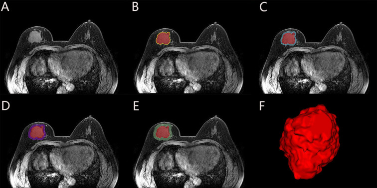
A total of 214 patients were randomly divided into a training set (n=149) and a test set (n=65) in a ratio of 7:3. Radiomics features were extracted from intratumoral region and 2-mm, 4-mm, 6-mm, 8-mm peritumoral regions on DCE-MRI images, and selected the optimal peritumoral region. The intratumoral radiomics model (IRM), 2-mm, 4-mm, 6-mm, 8-mm peritumoral radiomics model (PRM), the combined intra- and the optimal peritumoral radiomics model (CIPRM) were constructed based on five machine learning algorithms, and then the radiomics scores (Rad-score) were obtained. Independent risk factors for clinical-radiological features were obtained by univariate and multivariate logistic regression analysis, and clinical model (CM) was constructed. Finally, the CIPRM Rad-score combined with clinical-radiological factors was used to construct a NM. The performance of different models were evaluated by receiver operating characteristic curve (ROC) analysis, calibration curve analysis, and decision curve analysis (DCA).

RESULTS

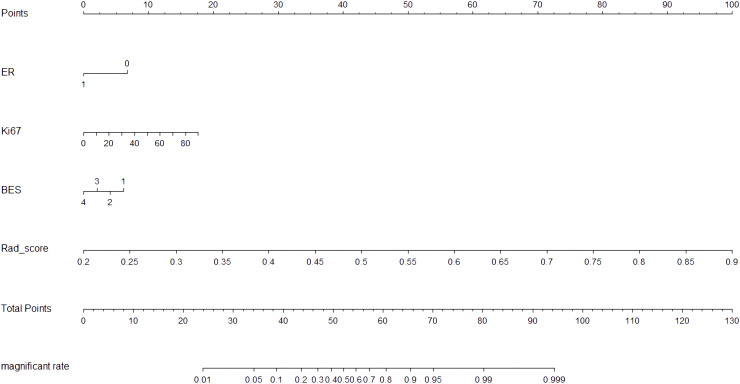
In our study, the 6-mm peritumoral size was considered to be the optimal peritumoral region. The CM is constructed based on three independent risk factors: estrogen receptor (ER), Ki-67, and breast edema score (BES). Incorporating ER, Ki-67, BES, and CIPRM Rad-score (combined intra- and 6-mm peritumoral) into the nomogram achieved a reliable predictive performance. And the area under the curve (AUC), sensitivity, specificity, and accuracy of the NM was 0.911, 0.848, 0.831, 0.826 for the training set and 0.897, 0.893, 0.784, 0.815 for the test set, respectively.

CONCLUSION

The NM has a good value for early prediction of pCR after NAC in breast cancer patients.

摘要

目的

本研究旨在创建一种列线图模型(NM),该模型将临床放射学因素与从治疗前动态对比增强磁共振成像(DCE-MRI)图像中提取的肿瘤内和瘤周区域的放射组学特征相结合,以建立一种可靠的方法来早期预测乳腺癌患者新辅助化疗(NAC)后的病理完全缓解(pCR)。

方法

总共214例患者以7:3的比例随机分为训练集(n = 149)和测试集(n = 65)。从DCE-MRI图像上的肿瘤内区域以及瘤周2毫米、4毫米、6毫米、8毫米区域提取放射组学特征,并选择最佳瘤周区域。基于五种机器学习算法构建肿瘤内放射组学模型(IRM)、2毫米、4毫米、6毫米、8毫米瘤周放射组学模型(PRM)、肿瘤内与最佳瘤周联合放射组学模型(CIPRM),然后获得放射组学评分(Rad-score)。通过单因素和多因素逻辑回归分析获得临床放射学特征的独立危险因素,并构建临床模型(CM)。最后,将CIPRM Rad-score与临床放射学因素相结合构建NM。通过受试者工作特征曲线(ROC)分析、校准曲线分析和决策曲线分析(DCA)评估不同模型的性能。

结果

在我们的研究中,6毫米的瘤周大小被认为是最佳瘤周区域。CM基于三个独立危险因素构建:雌激素受体(ER)、Ki-67和乳腺水肿评分(BES)。将ER、Ki-67、BES和CIPRM Rad-score(肿瘤内与6毫米瘤周联合)纳入列线图可实现可靠的预测性能。训练集的列线图曲线下面积(AUC)、敏感性、特异性和准确性分别为0.911、0.848、0.831、0.826,测试集分别为0.897、0.893、0.784、0.815。

结论

该列线图对乳腺癌患者NAC后pCR的早期预测具有良好价值。

https://cdn.ncbi.nlm.nih.gov/pmc/blobs/7326/12174392/9c58da894457/fonc-15-1561599-g007.jpg
https://cdn.ncbi.nlm.nih.gov/pmc/blobs/7326/12174392/3c276b08ab0a/fonc-15-1561599-g001.jpg
https://cdn.ncbi.nlm.nih.gov/pmc/blobs/7326/12174392/848db2a5fbb6/fonc-15-1561599-g002.jpg
https://cdn.ncbi.nlm.nih.gov/pmc/blobs/7326/12174392/007e9d213557/fonc-15-1561599-g003.jpg
https://cdn.ncbi.nlm.nih.gov/pmc/blobs/7326/12174392/c88a97923bda/fonc-15-1561599-g004.jpg
https://cdn.ncbi.nlm.nih.gov/pmc/blobs/7326/12174392/be2f0ae32608/fonc-15-1561599-g005.jpg
https://cdn.ncbi.nlm.nih.gov/pmc/blobs/7326/12174392/376bd14834ac/fonc-15-1561599-g006.jpg
https://cdn.ncbi.nlm.nih.gov/pmc/blobs/7326/12174392/9c58da894457/fonc-15-1561599-g007.jpg

相似文献

1
Intra- and peritumoral radiomics nomogram based on DCE-MRI for the early prediction of pathological complete response to neoadjuvant chemotherapy in breast cancer.基于动态对比增强磁共振成像(DCE-MRI)的瘤内及瘤周影像组学列线图用于早期预测乳腺癌新辅助化疗的病理完全缓解
Front Oncol. 2025 Jun 4;15:1561599. doi: 10.3389/fonc.2025.1561599. eCollection 2025.
2
Nomogram based on radiomics and CT features for predicting visceral pleural invasion of invasive adenocarcinoma ≤ 2 cm: A multicenter study.基于影像组学和CT特征的列线图预测直径≤2 cm浸润性腺癌脏层胸膜侵犯:一项多中心研究
Eur J Radiol. 2025 Jun 12;190:112227. doi: 10.1016/j.ejrad.2025.112227.
3
Peritumoral Radiomic Features on CT for Differential Diagnosis in Small-Cell Lung Cancer: Potential for Surgical Decision-Making.CT上小细胞肺癌鉴别诊断的瘤周放射组学特征:手术决策的潜力
Cancer Control. 2025 Jan-Dec;32:10732748251351754. doi: 10.1177/10732748251351754. Epub 2025 Jun 16.
4
Combination of ultrasound-based radiomics and deep learning with clinical data to predict response in breast cancer patients treated with neoadjuvant chemotherapy.基于超声的影像组学和深度学习与临床数据相结合,以预测接受新辅助化疗的乳腺癌患者的反应。
Front Oncol. 2025 Jun 5;15:1525285. doi: 10.3389/fonc.2025.1525285. eCollection 2025.
5
Delta-radiomics features combined with haematological index predict pathological complete response after neoadjuvant immunochemotherapy in resectable non-small cell lung cancer.Delta放射组学特征联合血液学指标预测可切除非小细胞肺癌新辅助免疫化疗后的病理完全缓解
Clin Radiol. 2025 Jul;86:106906. doi: 10.1016/j.crad.2025.106906. Epub 2025 Apr 7.
6
Dual-energy CT Radiomics Combined with Quantitative Parameters for Differentiating Lung Adenocarcinoma From Squamous Cell Carcinoma: A Dual-center Study.双能量CT影像组学联合定量参数鉴别肺腺癌与肺鳞癌:一项双中心研究
Acad Radiol. 2025 Mar;32(3):1675-1684. doi: 10.1016/j.acra.2024.09.024. Epub 2024 Sep 25.
7
Habitat-Based Radiomics for Revealing Tumor Heterogeneity and Predicting Residual Cancer Burden Classification in Breast Cancer.基于栖息地的放射组学在揭示乳腺癌肿瘤异质性及预测残留癌负荷分类中的应用
Clin Breast Cancer. 2025 Jul;25(5):444-454. doi: 10.1016/j.clbc.2025.01.014. Epub 2025 Feb 4.
8
A novel nomogram for predicting osteoporosis with low back pain among the patients in Wenshan Zhuang and Miao Autonomous Prefecture of China.中国文山壮族苗族自治州患者中用于预测伴有腰痛的骨质疏松症的新型列线图。
Front Endocrinol (Lausanne). 2025 Jun 5;16:1535163. doi: 10.3389/fendo.2025.1535163. eCollection 2025.
9
Development and validation of a Log odds of negative lymph nodes/T stage ratio-based prognostic model for gastric cancer.基于阴性淋巴结/肿瘤分期比值的胃癌对数优势预后模型的开发与验证
Front Oncol. 2025 Jun 3;15:1554270. doi: 10.3389/fonc.2025.1554270. eCollection 2025.
10
Transcriptome analysis and artificial intelligence for predicting lymph node metastasis of esophageal squamous cell carcinoma.用于预测食管鳞状细胞癌淋巴结转移的转录组分析与人工智能
J Thorac Dis. 2025 May 30;17(5):3283-3296. doi: 10.21037/jtd-2025-662. Epub 2025 May 28.

本文引用的文献

1
Machine learning radiomics based on intra and peri tumor PA/US images distinguish between luminal and non-luminal tumors in breast cancers.基于肿瘤内部及周边PA/US图像的机器学习放射组学可区分乳腺癌中的管腔型肿瘤和非管腔型肿瘤。
Photoacoustics. 2024 Sep 23;40:100653. doi: 10.1016/j.pacs.2024.100653. eCollection 2024 Dec.
2
Breast cancer statistics 2024.2024 年乳腺癌统计数据。
CA Cancer J Clin. 2024 Nov-Dec;74(6):477-495. doi: 10.3322/caac.21863. Epub 2024 Oct 1.
3
Delta Radiomics Based on MRI for Predicting Axillary Lymph Node Pathologic Complete Response After Neoadjuvant Chemotherapy in Breast Cancer Patients.
基于MRI的Delta放射组学预测乳腺癌患者新辅助化疗后腋窝淋巴结病理完全缓解
Acad Radiol. 2025 Jan;32(1):37-49. doi: 10.1016/j.acra.2024.07.052. Epub 2024 Sep 13.
4
Use of Pretreatment Perfusion MRI-based Intratumoral Heterogeneity to Predict Pathologic Response of Triple-Negative Breast Cancer to Neoadjuvant Chemoimmunotherapy.使用预处理灌注 MRI 评估肿瘤内异质性预测三阴性乳腺癌新辅助化疗免疫治疗的病理反应。
Radiology. 2024 Sep;312(3):e240575. doi: 10.1148/radiol.240575.
5
Utilizing multiclassifier radiomics analysis of ultrasound to predict high axillary lymph node tumour burden in node-positive breast cancer patients: a multicentre study.利用超声多分类放射组学分析预测腋窝淋巴结阳性乳腺癌患者高腋窝淋巴结肿瘤负荷:一项多中心研究。
Ann Med. 2024 Dec;56(1):2395061. doi: 10.1080/07853890.2024.2395061. Epub 2024 Aug 28.
6
Enhancing pathological complete response prediction in breast cancer: the role of dynamic characterization of DCE-MRI and its association with tumor heterogeneity.提高乳腺癌病理完全缓解预测:DCE-MRI 的动态特征及其与肿瘤异质性的相关性。
Breast Cancer Res. 2024 May 14;26(1):77. doi: 10.1186/s13058-024-01836-3.
7
Cancer statistics, 2024.2024年癌症统计数据。
CA Cancer J Clin. 2024 Jan-Feb;74(1):12-49. doi: 10.3322/caac.21820. Epub 2024 Jan 17.
8
Contrast-Enhanced Mammography Radiomics Analysis for Preoperative Prediction of Breast Cancer Molecular Subtypes.对比增强乳腺 X 线摄影影像组学分析在乳腺癌分子亚型术前预测中的应用。
Acad Radiol. 2024 Jun;31(6):2228-2238. doi: 10.1016/j.acra.2023.12.005. Epub 2023 Dec 23.
9
Development of an Intratumoral and Peritumoral Radiomics Nomogram Using Digital Breast Tomosynthesis for Preoperative Assessment of Lymphovascular Invasion in Invasive Breast Cancer.基于数字乳腺断层合成术的肿瘤内和肿瘤周放射组学列线图的建立,用于术前评估浸润性乳腺癌的淋巴管血管侵犯。
Acad Radiol. 2024 May;31(5):1748-1761. doi: 10.1016/j.acra.2023.11.010. Epub 2023 Dec 13.
10
Intratumoral and Peritumoral Radiomics Based on Preoperative MRI for Evaluation of Programmed Cell Death Ligand-1 Expression in Breast Cancer.基于术前 MRI 的肿瘤内和肿瘤周围放射组学分析预测乳腺癌程序性死亡配体-1 表达。
J Magn Reson Imaging. 2024 Aug;60(2):588-599. doi: 10.1002/jmri.29109. Epub 2023 Nov 2.