Patanè Vittorio, Atripaldi Umberto, Sansone Mario, Marinelli Luca, Del Tufo Sara, Arrichiello Gianluca, Ciardiello Davide, Selvaggi Francesco, Martinelli Erika, Reginelli Alfonso
Department of Precision Medicine, University of Campania "Luigi Vanvitelli", Piazza Luigi Miraglia 2, 80138, Naples, Italy.
Department of Electrical Engineering and Information Technology, University of Naples "Federico II", 80125, Naples, Italy.
Int J Colorectal Dis. 2025 Aug 8;40(1):174. doi: 10.1007/s00384-025-04969-9.
Preoperative T-staging in rectal cancer is essential for treatment planning, yet conventional MRI shows limited accuracy (~ 60-78). Our study investigates whether radiomic analysis of high-resolution T2-weighted MRI can non-invasively improve staging accuracy through a retrospective evaluation in a real-world surgical cohort.
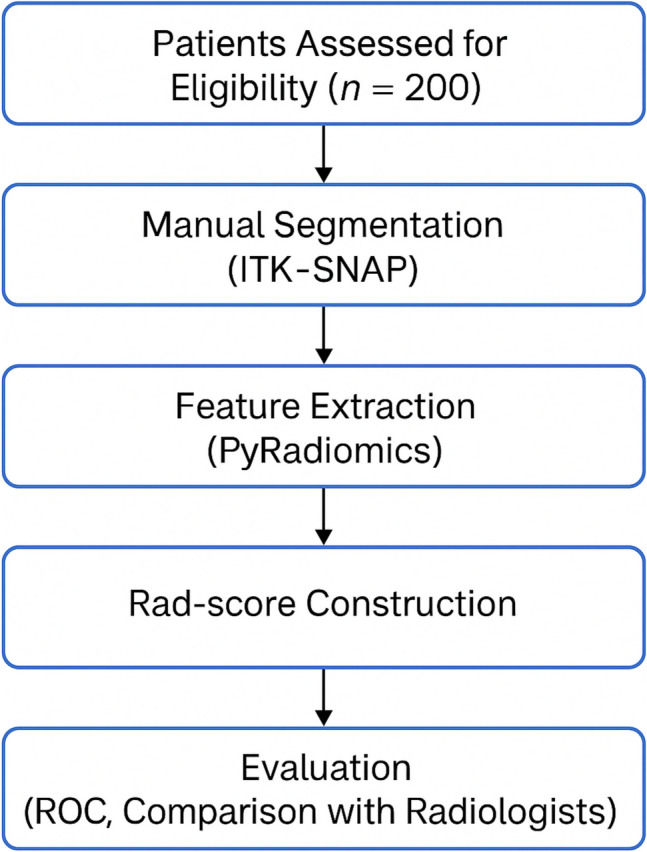
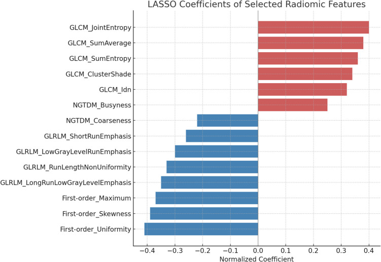
This single-center retrospective study included 200 patients (January 2024-April 2025) with pathologically confirmed rectal cancer, all undergoing preoperative high-resolution T2-weighted MRI within one week prior to curative surgery and no neoadjuvant therapy. Manual segmentation was performed using ITK‑SNAP, followed by extraction of 107 radiomic features via PyRadiomics. Feature selection employed mRMR and LASSO logistic regression, culminating in a Rad-score predictive model. Statistical performance was evaluated using ROC curves (AUC), accuracy, sensitivity, specificity, and Delong's test.
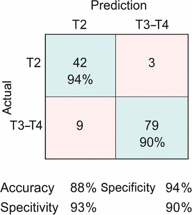
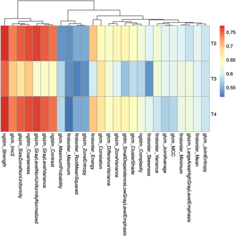
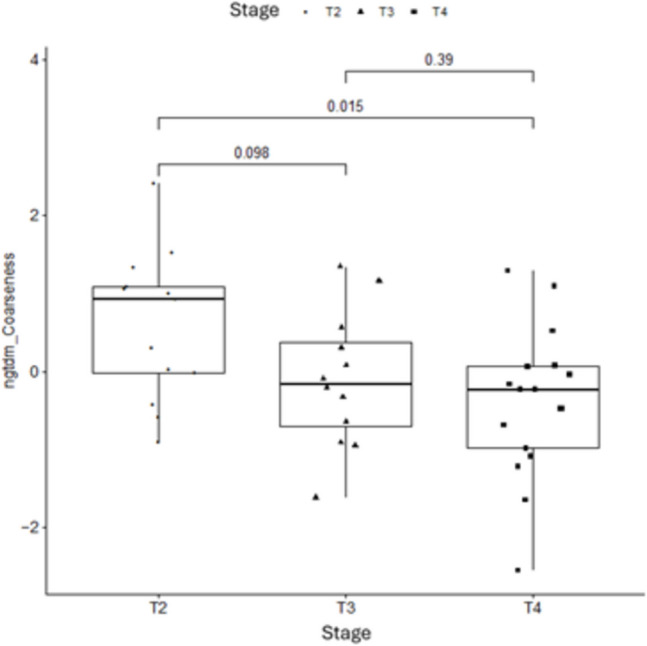
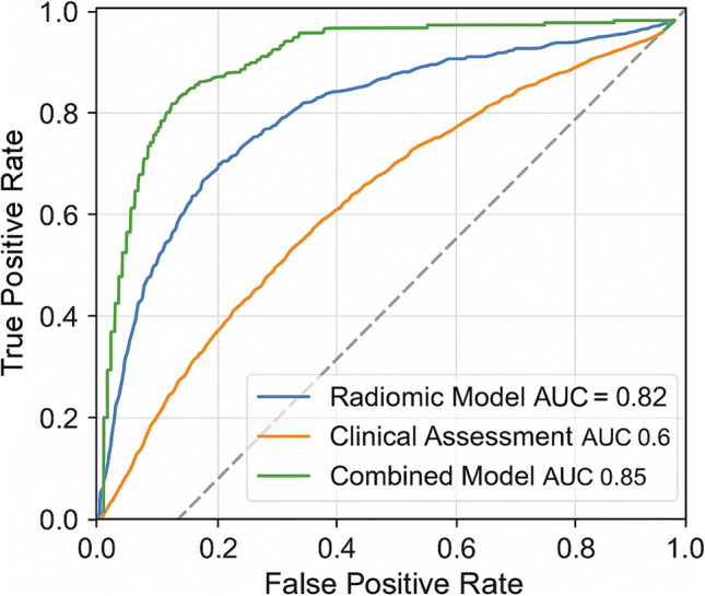
Among 200 patients, 95 were pathologically staged as T2 and 105 as T3-T4 (55 T3, 50 T4). After preprocessing, 26 radiomic features were retained; key features including ngtdm_contrast and ngtdm_coarseness showed AUC values > 0.70. The LASSO-based model achieved an AUC of 0.82 (95% CI: 0.75-0.89), with overall accuracy of 81%, sensitivity of 78%, and specificity of 84%.
Radiomic analysis of standard preoperative T2-weighted MRI provides a reliable, non-invasive method to predict rectal cancer T-stage. This approach has the potential to enhance staging accuracy and inform personalized surgical planning. Prospective multicenter validation is required for broader clinical implementation.
直肠癌术前T分期对于治疗方案的制定至关重要,但传统MRI的准确性有限(约60%-78%)。我们的研究通过对一个真实世界手术队列进行回顾性评估,探讨高分辨率T2加权MRI的放射组学分析能否无创地提高分期准确性。
这项单中心回顾性研究纳入了200例(2024年1月至2025年4月)经病理证实的直肠癌患者,所有患者均在根治性手术前一周内接受了术前高分辨率T2加权MRI检查,且未接受新辅助治疗。使用ITK-SNAP进行手动分割,随后通过PyRadiomics提取107个放射组学特征。特征选择采用mRMR和LASSO逻辑回归,最终建立了Rad评分预测模型。使用ROC曲线(AUC)、准确性、敏感性、特异性和德龙检验评估统计性能。
200例患者中,95例病理分期为T2,105例为T3-T4(55例T3,50例T4)。预处理后,保留了26个放射组学特征;包括ngtdm_contrast和ngtdm_coarseness在内的关键特征的AUC值>0.70。基于LASSO的模型AUC为0.82(95%CI:0.75-0.89),总体准确率为81%,敏感性为78%,特异性为84%。
标准术前T2加权MRI的放射组学分析提供了一种可靠的无创方法来预测直肠癌T分期。这种方法有可能提高分期准确性并为个性化手术规划提供依据。需要进行前瞻性多中心验证以实现更广泛的临床应用。