Lefol L A, Sodano A, Bawuah P, Zeitler J A, Verin J, Danede F, Willart J F, Siepmann J, Siepmann F
Univ. Lille, Inserm, CHU Lille, U1008, F-59000 Lille, France.
Univ. Cambridge, Department of Chemical Engineering and Biotechnology, Cambridge CB3 0AS, UK.
Int J Pharm X. 2025 Jul 23;10:100366. doi: 10.1016/j.ijpx.2025.100366. eCollection 2025 Dec.
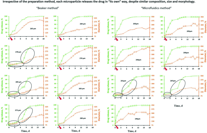
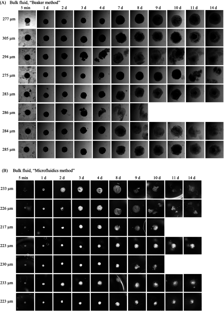
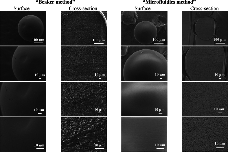
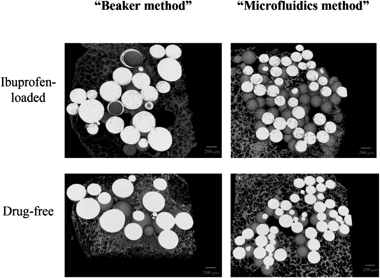
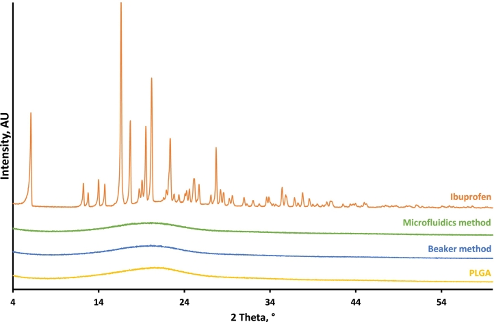
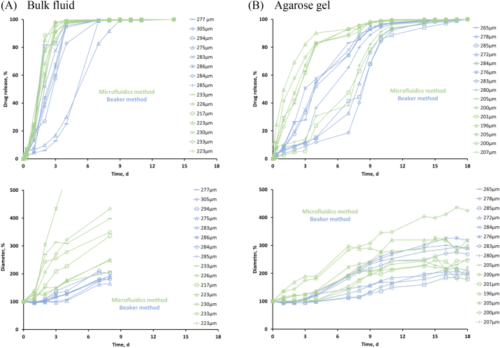
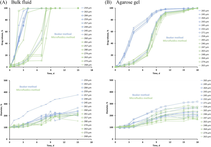
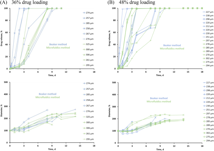
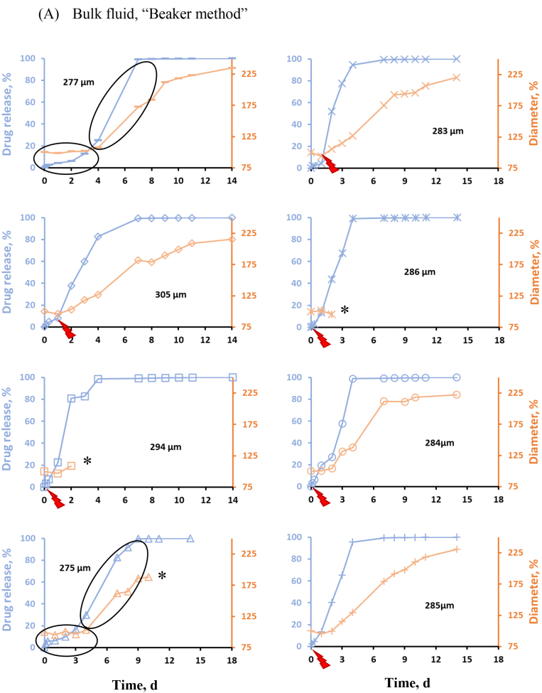
The aim of this study was to better understand the release mechanisms of poly(lactic--glycolic acid) (PLGA) microparticles prepared via emulsification - solvent extraction/evaporation using a "classical beaker" vs. a "microfluidics device". Ibuprofen-loaded microparticles were studied by optical microscopy, SEM, X-ray powder diffraction, X-ray μCT and drug release measurements from microparticles in well agitated phosphate buffer pH 7.4 or agarose gel (mimicking living tissue). The use of a microfluidics device facilitated the preparation of microparticles with a less broad size distribution. However, in addition to the microparticle size, the inner system structure was found to be also of utmost importance for the resulting drug release kinetics in this case. Interestingly, even microparticles with exhibited a . This was true, irrespective of the type of preparation method and experimental release set-up, and could be explained as follows: The investigated microparticles were characterized by a continuous inner pore network and an initially smooth & non-porous surface. Drug release set on as soon as: (i) the pore network got direct access to the release medium (e.g., due to a "weak point" in the PLGA surface layer), or (ii) substantial system swelling started (after a lag-time of several days). Importantly, microparticle had its own, specific structure, which determined "" to release the drug. Furthermore, the experimental conditions were found to be of key importance: The presence of a surrounding agarose gel protected the microparticles from damage caused by convective fluid flow, and hindered microparticle swelling, thus, slowing down drug release.
本研究的目的是更深入地了解通过乳化 - 溶剂萃取/蒸发法,使用“经典烧杯”与“微流控装置”制备聚乳酸 - 乙醇酸共聚物(PLGA)微粒的释放机制。通过光学显微镜、扫描电子显微镜(SEM)、X射线粉末衍射、X射线显微计算机断层扫描(X射线μCT)以及在pH 7.4的充分搅拌的磷酸盐缓冲液或琼脂糖凝胶(模拟活体组织)中对微粒进行药物释放测量,对载布洛芬微粒进行了研究。使用微流控装置有助于制备尺寸分布较窄的微粒。然而,在这种情况下,除了微粒尺寸外,内部系统结构对于最终的药物释放动力学也至关重要。有趣的是,即使是具有[此处原文缺失相关描述]的微粒也表现出[此处原文缺失相关描述]。无论制备方法的类型和实验释放设置如何,都是如此,其原因如下:所研究的微粒具有连续的内部孔隙网络和初始光滑且无孔的表面。药物释放一旦开始:(i)孔隙网络直接与释放介质接触(例如,由于PLGA表面层中的“弱点”),或(ii)在经过几天的滞后时间后开始大量系统溶胀。重要的是,每个微粒都有其自身特定的结构,该结构决定了[此处原文缺失相关描述]以释放药物。此外,发现实验条件至关重要:周围琼脂糖凝胶的存在保护微粒免受对流流体流动造成的损害,并阻碍微粒溶胀,从而减缓药物释放。