Chen Xianbo, Tao Xiaohong, Wang Jingyu
Department of Pediatrics, Wenling Maternal and Child Health Care Hospital, Wenling, Zhejiang, China.
Department of Neurosurgery, the Second Affiliated Hospital, Zhejiang University School of Medicine, Hangzhou, Zhejiang, China.
FASEB J. 2025 Aug 31;39(16):e70929. doi: 10.1096/fj.202402891RR.
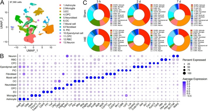
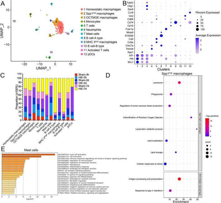
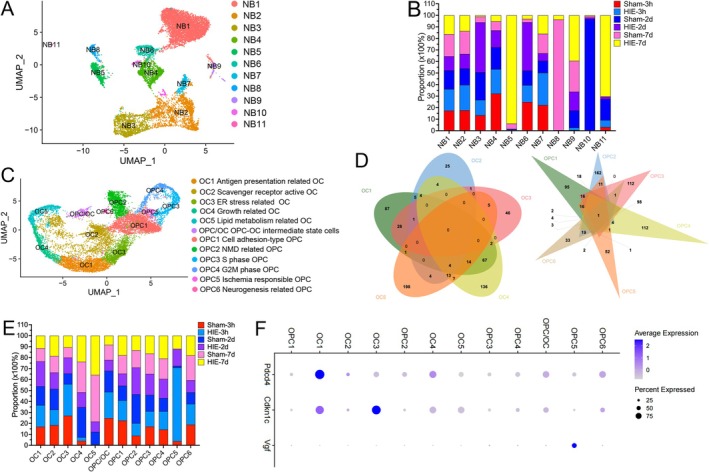
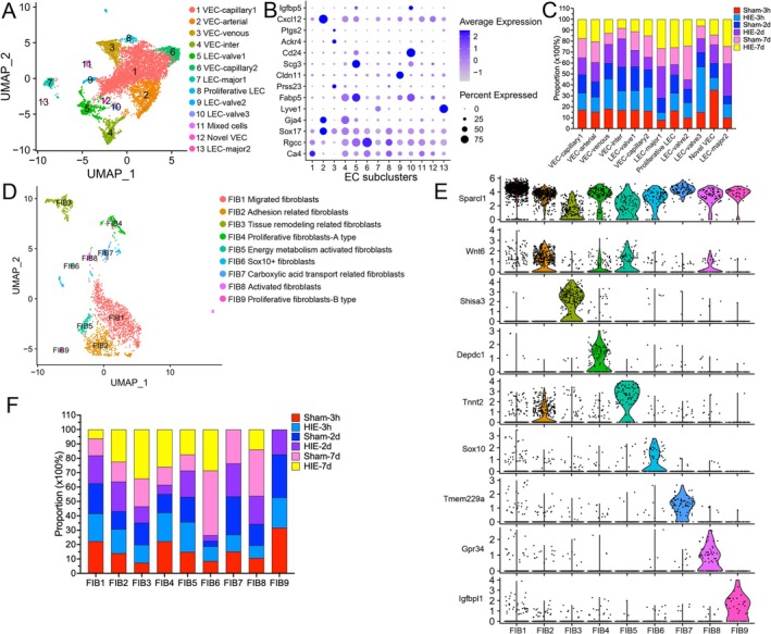
Neonatal hypoxic-ischemic encephalopathy (HIE) is a severe neurological condition associated with high rates of mortality or long-term disability. Despite its clinical significance, the detailed cellular mechanisms underlying HIE remain unclear. Single-cell RNA sequencing (scRNA-seq) has emerged as a powerful tool for investigating cellular heterogeneity across development, aging, and disease processes. However, no scRNA-seq studies have yet addressed neonatal HIE. In this study, we employed scRNA-seq to examine cellular heterogeneity during neonatal HIE. We analyzed a total of 87 580 high-quality brain cells to identify transcriptional changes associated with HIE. In the hyperacute phase, we observed astrocytes in response to tumor necrosis factors, involvement of microglia in phagocytosis, Stat3-mediated ischemic responses in oligodendrocyte precursor cells, and an increase in senescent lymphatic endothelial cells. In the acute phase, astrocytes were activated and involved in gliogenesis, while microglia proliferated. Neuroblasts were affected by metal ions, and oligodendrocytes decreased. In the subacute phase, astrocytes involved in inflammation and antigen presentation, while inflammatory microglia highly expressing MHC II were induced by the IL27 and type I interferon pathways and expanded. Additionally, peripheral immune cells played vital roles in HIE. Specifically, neutrophils infiltrated and expanded throughout all phases post-HIE. Spp1 macrophages, T cells, and plasmacytoid dendritic cells increased during the acute and subacute phases, and B cells expanded during the subacute phase. This study offers deep insights into the molecular alterations of key cell types following HIE, elucidating the pathological processes involved. These findings have significant implications for developing effective clinical strategies for managing HIE.
新生儿缺氧缺血性脑病(HIE)是一种严重的神经系统疾病,与高死亡率或长期残疾率相关。尽管其具有临床意义,但HIE背后详细的细胞机制仍不清楚。单细胞RNA测序(scRNA-seq)已成为研究发育、衰老和疾病过程中细胞异质性的有力工具。然而,尚未有scRNA-seq研究涉及新生儿HIE。在本研究中,我们采用scRNA-seq来检查新生儿HIE期间的细胞异质性。我们总共分析了87580个高质量的脑细胞,以确定与HIE相关的转录变化。在超急性期,我们观察到星形胶质细胞对肿瘤坏死因子有反应,小胶质细胞参与吞噬作用,少突胶质前体细胞中Stat3介导的缺血反应,以及衰老的淋巴管内皮细胞增加。在急性期,星形胶质细胞被激活并参与神经胶质生成,而小胶质细胞增殖。神经母细胞受到金属离子的影响,少突胶质细胞减少。在亚急性期,星形胶质细胞参与炎症和抗原呈递,而高表达MHC II的炎性小胶质细胞由IL27和I型干扰素途径诱导并扩增。此外,外周免疫细胞在HIE中起重要作用。具体而言,中性粒细胞在HIE后的所有阶段均浸润并扩增。Spp1巨噬细胞、T细胞和浆细胞样树突状细胞在急性期和亚急性期增加,B细胞在亚急性期扩增。本研究深入洞察了HIE后关键细胞类型的分子改变,阐明了其中涉及的病理过程。这些发现对制定有效的HIE临床管理策略具有重要意义。