Saladrigas Catherine A, Speed Forest, Teel Alec, Zohrabi Mo, Miscles Eduardo J, Futia Gregory L, Baker Larry V, Zhang Ye, Kymissis Ioannis, Bright Victor M, Welle Cristin G, Restrepo Diego, Gopinath Juliet T, Gibson Emily A
Department of Electrical, Energy and Computer Engineering, University of Colorado Boulder, CO 80309,USA.
Department of Bioengineering, University of Colorado Anschutz Medical Campus, Aurora, CO 80045, USA.
bioRxiv. 2025 Aug 6:2025.08.04.668551. doi: 10.1101/2025.08.04.668551.
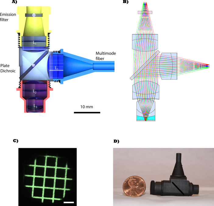
Functional imaging in freely moving animals with genetically encoded voltage indicators (GEVIs) will open new capabilities for neuroscientists to study the behavioral relevance of neural activity with high spatial and temporal precision. However, miniaturization of an imaging system with sufficient collection efficiency to resolve the small changes in fluorescence yield from voltage spikes, as well as development of efficient image sensors that are sufficiently fast to capture them, has proven challenging. We present a miniaturized microscope designed for voltage imaging, with a numerical aperture of 0.6, 250 μm field of view and 1.3 mm working distance that weighs 16.4 g. We show it is capable of imaging in vivo voltage spikes from Voltron2 with a spike peak-to-noise ratio >3 at a framerate of 530 Hz.
利用基因编码电压指示器(GEVIs)对自由活动动物进行功能成像,将为神经科学家开辟新的能力,使其能够以高空间和时间精度研究神经活动与行为的相关性。然而,要将成像系统小型化,使其具有足够的采集效率以分辨电压尖峰引起的荧光产率的微小变化,同时开发出足够快速以捕捉这些变化的高效图像传感器,已被证明具有挑战性。我们展示了一种专为电压成像设计的小型显微镜,其数值孔径为0.6,视野为250μm,工作距离为1.3mm,重量为16.4g。我们表明它能够在530Hz的帧率下对Voltron2的体内电压尖峰进行成像,尖峰峰噪比>3。