Zhang Zelin, Yang Qingsong, Shiradkar Rakesh, Mirtti Tuomas, Azamat Sena, Xuan Kai, Xu Jun, Madabhushi Anant
Wallace H. Coulter Department of Biomedical Engineering, Georgia Institute of Technology and Emory University, Atlanta, Georgia, USA.
Changhai Hospital, Shanghai, China.
Med Phys. 2025 Aug;52(8):e18053. doi: 10.1002/mp.18053.
Benign prostatic hyperplasia (BPH) and prostate cancer (PCa) share overlapping characteristics on magnetic resonance imaging (MRI), confounding the diagnosis and detection of PCa. There is thus a clinical need to accurately differentiate BPH-Only from BPH-PCa to prevent overdiagnosis and unnecessary biopsies. Although BPH and PCa may share overlapping features, they are distinct clinical entities. Previous evidence suggests that prostate peripheral zone (PZ) and transition zone (TZ) volumes on MRI are differentially associated in patients with BPH-PCa versus those with BPH-Only.
To develop and validate the ratio of machine learning derived PZ and TZ volumes on T2-weighted (T2W) MRI as an imaging biomarker to distinguish BPH-PCa and BPH-Only.
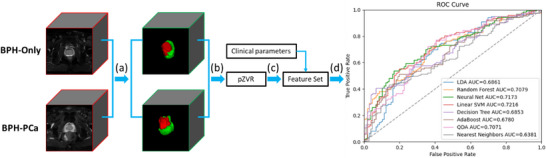
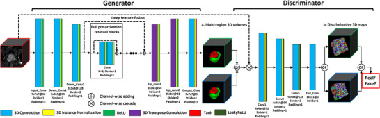
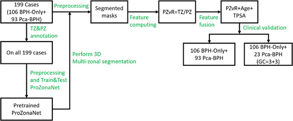
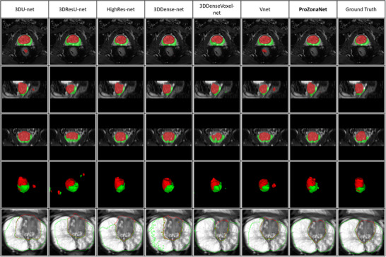
In this single-center, retrospective study, we identified N = 199 patients (106 BPH-Only and 93 with both BPH-PCa) who underwent a three Tesla multi-parametric MRI before systematic biopsy. A radiologist and a urologist jointly annotated PZ and TZ regions of interest on T2W, involving all 199 cases. We presented and trained a 3D conditional generative adversarial network (cGAN)-based prostate zone volume segmentation model (ProZonaNet) to segment 3D prostate TZ, PZ volumes on T2W MRI. We used 139 cases (with 7× data augmentation, yielding 973 training volumes) and an independent test set of 60 cases to train and evaluate ProZonaNet. ProZonaNet was optimized in terms of dice similarity coefficient (DSC). We then computed prostate zonal volume ratio (pZVR = TZ/PZ) from both ProZonaNet segmentations and ground-truth annotations on all 199 cases, evaluating agreement using Concordance Correlation Coefficient (CCC). The pZVR biomarker was assessed for its ability to distinguish BPH-PCa from BPH-Only. Univariate and multivariate analyses were performed to evaluate the independent effect of pZVR over clinical parameters.
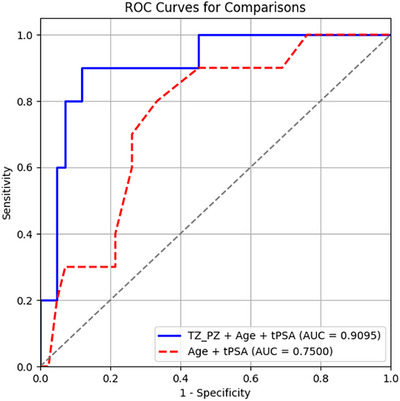
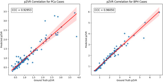
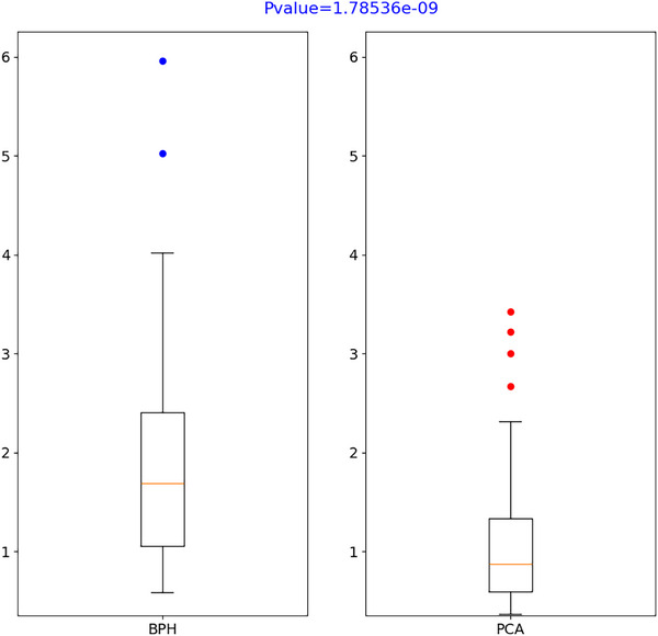
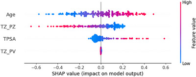
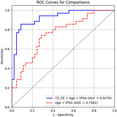
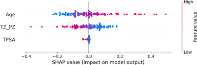
ProZonaNet achieved a mean mDICE of 92.5% on the independent test set (N = 60), outperforming state-of-the-art 3D segmentation models. The computed pZVR showed high agreement with ground-truth annotations, with CCC values of 0.960 for BPH-Only and 0.930 for BPH-PCa cases. The pZVR computed using ProZonaNet, along with two other clinical parameters, including age and prostate-specific antigen, improved the AUC from 0.758 to 0.927 in distinguishing between BPH-Only and BPH-PCa. At the same time, on a subset of low-grade prostate cancer cases (106 BPH-Only and 23 BPH-PCa with Gleason Score = 3+3), the integrated pZVR model improved the AUC from 0.750 to 0.910 in distinguishing between patients with BPH-Only versus BPH-PCa. On both univariate and multivariate analyses, pZVR demonstrated significant discrimination between patients with BPH-Only versus BPH-PCa.
We demonstrated that the prostate zonal volume ratio computed with our ProZonaNet can be used to differentiate benign prostatic hyperplasia from prostate cancer on MRI. These results demonstrate the feasibility of non-invasive diagnosis of BPH-PCa, potentially aiding in the ability to distinguish PCa from benign cancer confounders such as BPH-Only.
良性前列腺增生(BPH)和前列腺癌(PCa)在磁共振成像(MRI)上具有重叠特征,这使得PCa的诊断和检测变得复杂。因此,临床上需要准确区分单纯BPH和BPH合并PCa,以防止过度诊断和不必要的活检。虽然BPH和PCa可能有重叠特征,但它们是不同的临床实体。先前的证据表明,在BPH合并PCa患者与单纯BPH患者中,MRI上前列腺外周带(PZ)和移行带(TZ)的体积存在差异关联。
开发并验证基于机器学习得出的T2加权(T2W)MRI上PZ和TZ体积之比作为一种成像生物标志物,以区分BPH合并PCa和单纯BPH。
在这项单中心回顾性研究中,我们纳入了199例患者(106例单纯BPH和93例BPH合并PCa),这些患者在进行系统活检前接受了3特斯拉多参数MRI检查。一名放射科医生和一名泌尿科医生共同在T2W图像上标注了199例患者的PZ和TZ感兴趣区域。我们展示并训练了一个基于3D条件生成对抗网络(cGAN)的前列腺区域体积分割模型(ProZonaNet),用于分割T2W MRI上的3D前列腺TZ、PZ体积。我们使用139例病例(经过7倍数据增强,产生973个训练体积)和一个包含60例病例的独立测试集来训练和评估ProZonaNet。ProZonaNet在骰子相似系数(DSC)方面进行了优化。然后,我们根据ProZonaNet分割结果和所有199例病例的真实标注计算前列腺区域体积比(pZVR = TZ/PZ),使用一致性相关系数(CCC)评估一致性。评估pZVR生物标志物区分BPH合并PCa和单纯BPH的能力。进行单因素和多因素分析以评估pZVR相对于临床参数的独立效应。
ProZonaNet在独立测试集(N = 60)上的平均mDICE为92.5%,优于现有最先进的3D分割模型。计算得出的pZVR与真实标注高度一致,单纯BPH病例的CCC值为0.960,BPH合并PCa病例的CCC值为0.930。使用ProZonaNet计算的pZVR以及另外两个临床参数,包括年龄和前列腺特异性抗原,在区分单纯BPH和BPH合并PCa时,将曲线下面积(AUC)从0.758提高到了0.927。同时,在一部分低级别前列腺癌病例(106例单纯BPH和23例Gleason评分为3 + 3的BPH合并PCa)中,综合pZVR模型在区分单纯BPH和BPH合并PCa患者时,将AUC从0.750提高到了0.910。在单因素和多因素分析中,pZVR在单纯BPH和BPH合并PCa患者之间均显示出显著的区分能力。
我们证明,使用我们的ProZonaNet计算得出的前列腺区域体积比可用于在MRI上区分良性前列腺增生和前列腺癌。这些结果证明了无创诊断BPH合并PCa的可行性,可能有助于将PCa与良性癌症混淆因素(如单纯BPH)区分开来。