Bogdanovic Ivan, Ilic Rosanda, Kostic Aleksandar, Miljkovic Aleksandar, Milisavljevic Filip, Janjic Marija M, Bjelobaba Ivana M, Savic Danijela, Bascarevic Vladimir
University Clinical Center of Serbia, Clinic for Neurosurgery, 11000 Belgrade, Serbia.
School of Medicine, University of Belgrade, 11000 Belgrade, Serbia.
Diagnostics (Basel). 2025 Jul 22;15(15):1836. doi: 10.3390/diagnostics15151836.
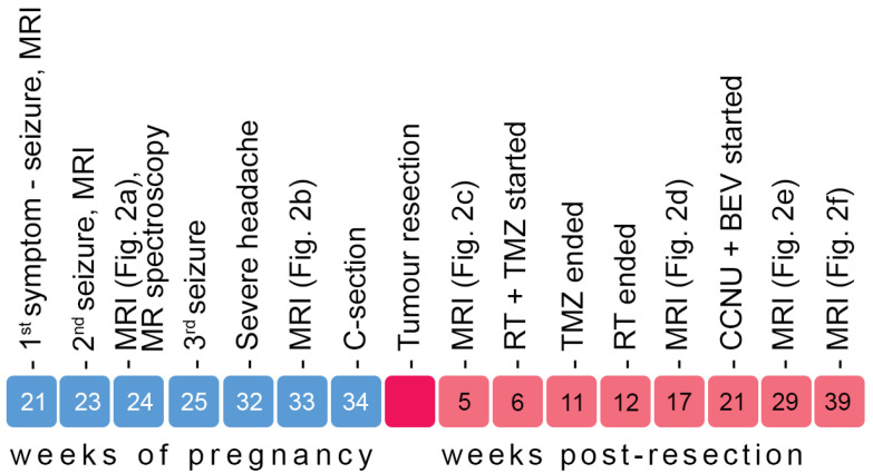
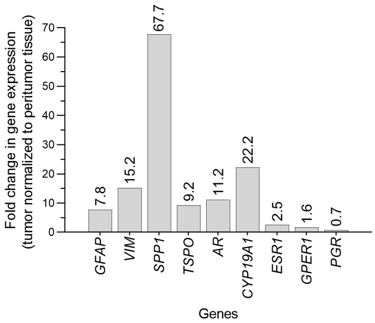
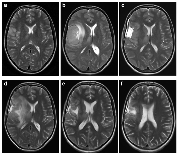
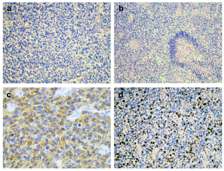
Gliomas diagnosed during pregnancy are rare, and there are no established guidelines for their management. Effective treatment requires a multidisciplinary approach to balance maternal health and pregnancy preservation. We here present a case of rapidly progressing glioma in a 33-year-old pregnant woman. The patient initially presented with a generalized tonic-clonic seizure at 21 weeks' gestation. Imaging revealed a tumor in the right cerebral lobe, involving both cortical and subcortical structures, while magnetic resonance spectroscopy suggested a low-grade glioma. The patient remained clinically stable for two months but then developed severe headaches; MRI showed a worsening mass effect. At 34 weeks' gestation, an emergency and premature caesarean section was performed under general anesthesia. The patient then underwent a craniotomy for maximal tumor resection, which was histologically and molecularly diagnosed as IDH wild-type glioblastoma (GB). Using qPCR, we found that the GB tissue showed upregulated expression of genes involved in cell structure (, ) and immune response (, ), as well as increased expression of genes related to potential hormone response (, , , ). After surgery, the patient showed resistance to Stupp protocol therapy, which was substituted with lomustine and bevacizumab combination therapy. This case illustrates that glioma may progress rapidly during pregnancy, but a favorable obstetric outcome is achievable. Management of similar cases should respect both the need for timely treatment and the patient's informed decision.
孕期诊断出的胶质瘤很罕见,目前尚无既定的治疗指南。有效的治疗需要多学科方法来平衡母体健康和保留妊娠。我们在此报告一例33岁孕妇快速进展性胶质瘤的病例。患者最初在妊娠21周时出现全身强直阵挛性发作。影像学检查显示右侧脑叶有一个肿瘤,累及皮质和皮质下结构,而磁共振波谱提示为低级别胶质瘤。患者临床稳定两个月,但随后出现严重头痛;磁共振成像显示占位效应恶化。妊娠34周时,在全身麻醉下进行了急诊剖宫产。随后患者接受了开颅手术以最大程度切除肿瘤,经组织学和分子学诊断为异柠檬酸脱氢酶(IDH)野生型胶质母细胞瘤(GB)。使用定量聚合酶链反应(qPCR),我们发现GB组织中参与细胞结构(……)和免疫反应(……)的基因表达上调,以及与潜在激素反应相关的基因(……、……、……、……)表达增加。术后,患者对Stupp方案治疗耐药,改用洛莫司汀和贝伐单抗联合治疗。该病例表明,胶质瘤在孕期可能快速进展,但可实现良好的产科结局。类似病例的管理应兼顾及时治疗的需求和患者的知情决定。