Li Jian, Xu Ruiping, Wang Guan, Su Yanhua, Chen Yaoxing, Cao Jing
Anhui Province Key Laboratory of Embryo Development and Reproductive Regulation, College of Biology and Food Engineering, Fuyang Normal University, Yingzhou, Fuyang 236037, China.
Laboratory of Anatomy of Domestic Animals, State Key Laboratory of Veterinary Public Health and Safety, College of Veterinary Medicine, China Agricultural University, Haidian, Beijing 100193, China.
Toxics. 2025 Aug 9;13(8):672. doi: 10.3390/toxics13080672.
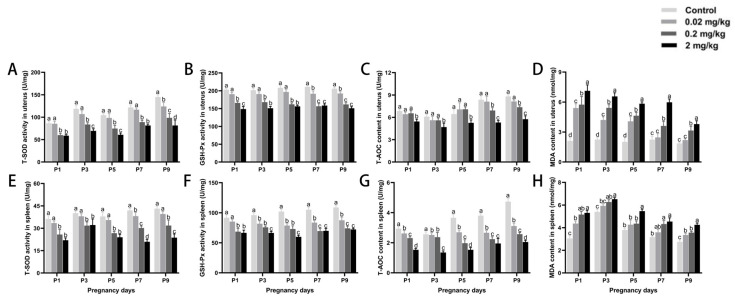
Due to the growing environmental burden of endocrine-disrupting chemicals (EDCs), there is an increasing concern regarding the reproductive hazards posed by synthetic estrogens, particularly diethylstilbestrol (DES). However, the precise mechanisms by which DES disrupts uterine endocrine function and immune homeostasis leading to pregnancy failure remain unclear. Given that wild rodents serve as key reservoirs for zoonotic diseases such as plague, reproductive interventions targeting their pregnancy processes hold significant ecological implications for disease control. In this study, female mice in estrus were randomly divided into four experimental groups, receiving DES at doses of 0 (control), 0.02 (low), 0.2 (medium), and 2 mg/kg (high), respectively. For five consecutive days, mice were injected subcutaneously on a daily basis, with the goal of examining DES-related alterations in hormone secretion and local immune responses within the uterus and spleen. It was found that high-dose DES treatment significantly increased maternal body weight and spleen weight during early pregnancy ( < 0.05). Meanwhile, reproductive function declined progressively with increasing doses, as indicated by decreased ovarian and uterine weights, fewer embryos, and extended estrous cycle duration ( < 0.05). Hematoxylin and eosin staining revealed that high-dose DES markedly reduced uterine gland density at day P5, accompanied by epithelial vacuolar degeneration and nuclear pyknosis. The proportion of uterine glands relative to total uterine area also decreased significantly with increasing DES doses. Moreover, DES inhibited lymphocyte proliferation in both the uterus and spleen in a dose-dependent fashion, with ConA- and LPS-induced proliferation rates decreasing by 0.78-30.70% and 1.91-18.20%, respectively ( < 0.05). The proinflammatory cytokine IL-2 was significantly elevated by DES, whereas the anti-inflammatory cytokine IL-4 showed a notable decrease ( < 0.05). DES administration notably decreased uterine expression of proliferating cell nuclear antigen. In contrast, the numbers of B-cell lymphoma 2- and Bcl-2-associated X protein-positive cells rose, along with upregulated levels of inducible nitric oxide synthase. Furthermore, DES impaired antioxidant defenses in both the uterus and spleen, evidenced by the decreased activities of superoxide dismutase and glutathione peroxidase, reduced total antioxidant capacity, and elevated malondialdehyde levels. This study elucidates the multifaceted mechanisms by which DES impairs the early gestational reproductive environment, filling a critical knowledge gap regarding its interference with the uterus-immune axis, and expands the current understanding of the ecotoxicological impacts of endocrine-disrupting chemicals.
由于内分泌干扰化学物质(EDCs)对环境造成的负担日益加重,人们越来越关注合成雌激素,尤其是己烯雌酚(DES)所带来的生殖危害。然而,DES破坏子宫内分泌功能和免疫稳态导致妊娠失败的确切机制仍不清楚。鉴于野生啮齿动物是鼠疫等人畜共患病的主要宿主,针对其妊娠过程的生殖干预对疾病控制具有重要的生态意义。在本研究中,处于发情期的雌性小鼠被随机分为四个实验组,分别接受剂量为0(对照)、0.02(低)、0.2(中)和2mg/kg(高)的DES。连续五天,每天对小鼠进行皮下注射,目的是研究DES对子宫和脾脏内激素分泌及局部免疫反应的相关改变。结果发现,高剂量DES处理显著增加了妊娠早期母体体重和脾脏重量(<0.05)。同时,随着剂量增加,生殖功能逐渐下降,表现为卵巢和子宫重量减轻、胚胎数量减少以及发情周期延长(<0.05)。苏木精-伊红染色显示,高剂量DES在妊娠第5天显著降低了子宫腺密度,伴有上皮细胞空泡变性和核固缩。子宫腺相对于子宫总面积的比例也随着DES剂量增加而显著降低。此外,DES以剂量依赖的方式抑制子宫和脾脏中的淋巴细胞增殖,伴刀豆蛋白A和脂多糖诱导的增殖率分别降低0.78 - 30.70%和1.91 - 18.20%(<0.05)。促炎细胞因子IL - 2被DES显著升高,而抗炎细胞因子IL - 4则显著降低(<0.05)。给予DES显著降低了子宫中增殖细胞核抗原的表达。相反,B细胞淋巴瘤2和Bcl - 2相关X蛋白阳性细胞数量增加,同时诱导型一氧化氮合酶水平上调。此外,DES损害了子宫和脾脏中的抗氧化防御能力,表现为超氧化物歧化酶和谷胱甘肽过氧化物酶活性降低、总抗氧化能力下降以及丙二醛水平升高。本研究阐明了DES损害早期妊娠生殖环境的多方面机制,填补了其对子宫 - 免疫轴干扰方面的关键知识空白,并扩展了当前对内分泌干扰化学物质生态毒理学影响的理解。