Uddin Md Imam, Jamal Sara, Penn John S
Vanderbilt University School of Medicine.
Res Sq. 2025 Aug 21:rs.3.rs-7247191. doi: 10.21203/rs.3.rs-7247191/v1.
Retinal hypoxia may contribute to the development of preretinal neovascularization in patients with retinopathy of prematurity (ROP). Ciliary bodies compensate oxygen delivery to the retina, and the levels of hypoxia may vary across the peripheral avascular area in ROP. In this study, we have investigated a direct method for imaging gradient levels of retinal hypoxia at the peripheral avascular retina using a model ROP.
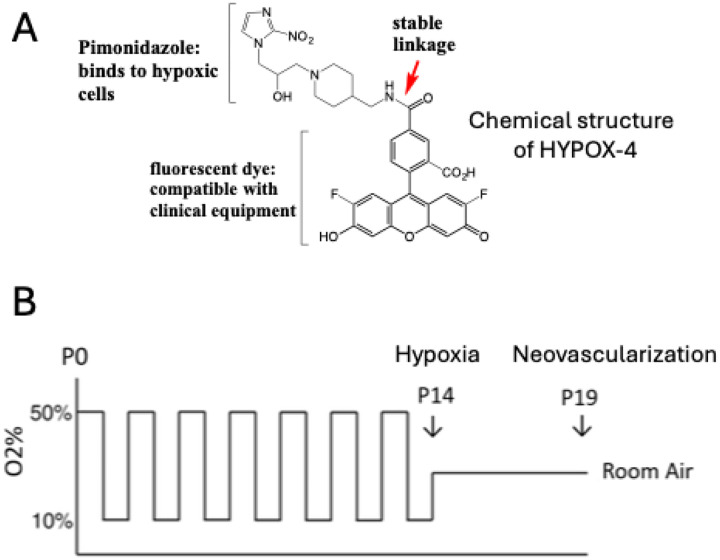
The rat 50/10 oxygen-induced retinopathy (OIR) model was generated by exposing the newly born Brown-Norway rat pups to a 24 hours alternate cycles of 50% and 10% oxygen for 14 days. We also confirmed the development of neovascularization in this model. HYPOX4 was used as a direct method for imaging gradient levels of retinal hypoxia at the peripheral avascular retina. A separate group of rat OIR pups were used to confirm gradient levels of retinal hypoxia using pimonidazole immunostaining. Gradient levels of retinal hypoxia was analyzed using ImageJ software from fluorescence intensities of HYPOX-4 and Pimonidazole immunostaining.
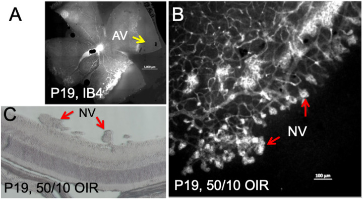
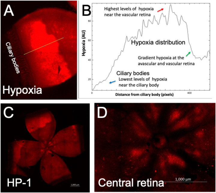
Retinal hypoxia was observed in the peripheral avascular retinas in rat OIR. Based on fluorescence intensity measurements, retinal hypoxia was at minimal levels near the ciliary bodies. Retinal hypoxia was at its maximum levels towards the avascular-vascular transition zones. Interestingly, we observed hemiretinal avascular retina temporal to the optic nerve in this OIR model, similar to human ROP retinas. In the retinal cross-section, hypoxia was not detectable near the ora serrata in rat OIR may be due to oxygen delivery by the ciliary bodies. Both pimonidazole and HYPOX-4 showed similar patterns of retinal hypoxia at the peripheral avascular retina in this model. As expected, preretinal neovascularization was observed at the avascular-vascular transition zones arising from the existing retinal vascular structures in this OIR model in Brown-Norway rats.
In this study, we have characterized gradient levels of retinal hypoxia in the rat model of 50/10 OIR using a direct method from HYPOX-4 fluorescence. We observed minimal levels of retinal hypoxia near the ciliary bodies in this model and increased towards the avascular-vascular transition zones. In addition, we observed that the central vascularized retina remains gradient hypoxic in this model which could be detected using HYPOX-4. This study may clarify our understanding of retinal hypoxia in the ROP patient at the peripheral retinas.
视网膜缺氧可能促成早产儿视网膜病变(ROP)患者视网膜前新生血管的形成。睫状体可代偿性地向视网膜输送氧气,且ROP患者外周无血管区的缺氧水平可能存在差异。在本研究中,我们利用ROP模型研究了一种直接成像外周无血管视网膜区域视网膜缺氧梯度水平的方法。
通过将新生的棕色挪威大鼠幼崽暴露于50%和10%氧气交替的环境中24小时,持续14天,建立大鼠50/10氧诱导性视网膜病变(OIR)模型。我们还证实了该模型中新生血管的形成。HYPOX4被用作直接成像外周无血管视网膜区域视网膜缺氧梯度水平的方法。另一组大鼠OIR幼崽用于通过匹莫硝唑免疫染色来确认视网膜缺氧的梯度水平。利用ImageJ软件根据HYPOX - 4和匹莫硝唑免疫染色的荧光强度分析视网膜缺氧的梯度水平。
在大鼠OIR模型的外周无血管视网膜中观察到了视网膜缺氧。根据荧光强度测量结果,睫状体附近的视网膜缺氧水平最低。朝向无血管 - 血管过渡区,视网膜缺氧水平最高。有趣的是,在该OIR模型中,我们观察到视神经颞侧半侧视网膜无血管,类似于人类ROP视网膜。在视网膜横切面上,大鼠OIR模型中锯齿缘附近未检测到缺氧,这可能是由于睫状体输送氧气所致。在该模型中,匹莫硝唑和HYPOX - 4在外周无血管视网膜中均显示出相似的视网膜缺氧模式。正如预期的那样,在棕色挪威大鼠的该OIR模型中,在由现有视网膜血管结构产生的无血管 - 血管过渡区观察到了视网膜前新生血管。
在本研究中,我们利用来自HYPOX - 4荧光的直接方法,对大鼠50/10 OIR模型中的视网膜缺氧梯度水平进行了特征描述。在该模型中,我们观察到睫状体附近的视网膜缺氧水平最低,且向无血管 - 血管过渡区增加。此外,我们观察到在该模型中,中央血管化视网膜也存在梯度性缺氧,可通过HYPOX - 4检测到。本研究可能会阐明我们对ROP患者外周视网膜缺氧的理解。