Akter Sumaya, Mirhosseiniardakani Soheila, Takekoshi Yuto, Qiu Liyan, Baker Kevin, Jiang Kevin, Lyon Mark, Hattori Mitsuharu, Chen Xuanmao
Department of Molecular, Cellular, and Biomedical Sciences; College of Life Sciences and Agriculture, University of New Hampshire, 46 College Road, Durham, NH 03824.
Department of Biomedical Science, Graduate School of Pharmaceutical Sciences, Nagoya City University, Nagoya, Aichi 467-8603, Japan.
bioRxiv. 2025 Aug 24:2025.08.24.672028. doi: 10.1101/2025.08.24.672028.
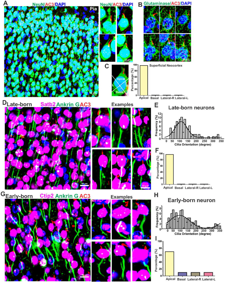
The primary cilia of pyramidal neurons in inside-out laminated regions orient predominantly toward the pial surface, reflecting reverse soma re-positioning during postnatal development. However, the mechanisms underlying the directional cilia orientation and reverse movement are unknown. Here we show that the primary cilia of pyramidal neurons are localized near the base of the apical dendrites and aligned on the nuclear side opposite to the axon initial segment. However, this pattern is not observed in atypical pyramidal neurons in the deep neocortex, excitatory neurons in non-laminated brain regions, interneurons, or astrocytes, where their cilia are irregularly positioned around the nuclei and lack preferred orientation. In Reelin-deficient mice (), the directional orientation and apical localization of cilia in late-born neocortical and CA1 neurons are partially impaired, because their initial impairments are partially corrected during late postnatal development, along with a realignment of apical-basal orientation. In contrast, loss of Reelin drastically disrupts the directional orientation of primary cilia in early-born neocortical neurons and principal neurons in the piriform cortex; consistently, their cilia and centrioles do not preferably localize to the base of the apical dendrites. Additionally, Reelin deficiency increases the cilia length of principal neurons in the cerebral cortex after P14 when WT cilia stabilize, but not in interneurons, astrocytes, or excitatory neurons in non-laminated regions. Together, Reelin controls the directional orientation, intracellular localization, and length of primary cilia in principal neurons in the cerebral cortex. These results underscore primary cilia as a key apical unit, particularly prominent in late-born neurons.
在由内向外分层区域的锥体神经元的初级纤毛主要朝向软膜表面定向,这反映了出生后发育过程中胞体的反向重新定位。然而,纤毛定向和反向运动的潜在机制尚不清楚。在这里,我们表明锥体神经元的初级纤毛位于顶端树突基部附近,并在与轴突起始段相对的核侧排列。然而,在深层新皮层的非典型锥体神经元、非分层脑区的兴奋性神经元、中间神经元或星形胶质细胞中未观察到这种模式,在这些细胞中,它们的纤毛围绕细胞核不规则定位且缺乏优先取向。在瑞连蛋白缺陷小鼠中,出生较晚的新皮层和CA1神经元中纤毛的定向和顶端定位部分受损,因为它们最初的损伤在出生后晚期发育过程中部分得到纠正,同时顶端 - 基部取向也重新排列。相比之下,瑞连蛋白的缺失会严重破坏出生较早的新皮层神经元和梨状皮层中的主要神经元的初级纤毛的定向;一致地,它们的纤毛和中心粒不会优先定位于顶端树突的基部。此外,当野生型纤毛稳定后,瑞连蛋白缺乏会增加P14后脑皮层主要神经元的纤毛长度,但不会增加非分层区域的中间神经元、星形胶质细胞或兴奋性神经元的纤毛长度。总之,瑞连蛋白控制着大脑皮层主要神经元中初级纤毛的定向、细胞内定位和长度。这些结果强调了初级纤毛作为关键的顶端单元,在出生较晚的神经元中尤为突出。