Taraschenko Olga, Arcot Jayagopal Lakshman, Mullane Audrina, Greenman Kyle, White Matthew, Ghonim Hesham, Lee Shelley, Zabad Rana Khalil, Jasinski Tracy, Uberti Mariano
Department of Neurological Sciences, Division of Epilepsy, University of Nebraska Medical Center, Omaha, NE, United States.
Department of Neurological Sciences, Division of Neuropsychology, University of Nebraska Medical Center, Omaha, NE, United States.
Front Neurol. 2025 Aug 14;16:1597928. doi: 10.3389/fneur.2025.1597928. eCollection 2025.
Autoimmune encephalitis (AE) is associated with severe cognitive disability. Brain metabolic dysfunction has been linked to encephalopathy in neurodegenerative disorders; however, its role in the development of cognitive loss in AE has not been studied. We hypothesized that cognitively impaired patients with AE will demonstrate altered brain metabolism and immune activation, and these measures will correlate with cognitive scores.
The hippocampal and cortical metabolites related to neuronal integrity, oxidative metabolism, and glial activation were assessed using single-voxel proton magnetic resonance spectroscopy (1H-MRS) in patients with AE, non-lesional temporal lobe epilepsy (TLE) and control subjects. Metabolite levels were correlated with neuropsychological test scores.
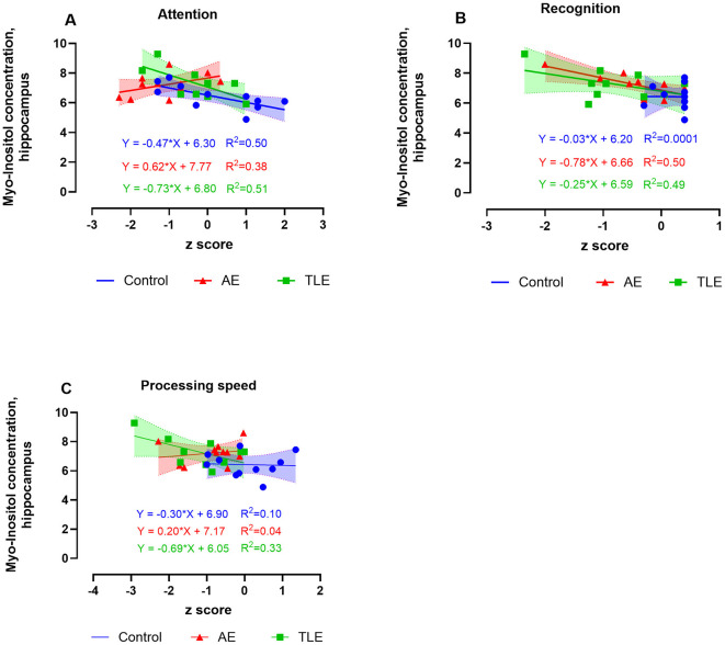
We recruited patients with post-acute AE ( = 12), non-lesional TLE ( = 12), and control subjects ( = 11). Subjective cognitive complaints were reported by 83.3% of AE and all TLE patients. AE patients had fewer seizures and used fewer anti-seizure medications than TLE patients ( = 0.04, -test and = 0.03, test). On neuropsychological testing, moderate and severe cognitive impairment was revealed in 58.3% of patients with AE and 41.6% of patients with TLE. Hippocampal myo-inositol (M-Ins) concentrations were higher in patients compared to control subjects, with a trend toward increase in AE and TLE relative to control ( = 0.046, ANOVA; = 0.09 and = 0.07 for AE and TLE vs. control, respectively; tests). The concentration of creatine (tCr) and total choline (tCho) were significantly higher in patients with TLE compared to the controls (tCr: = 0.007; tCh: = 0.04; tests). Elevated M-Ins in AE was associated with better attention but worse memory recognition scores ( = 0.38, = 0.04 and = 0.50, = 0.02, respectively); higher tCr levels correlated with faster processing speed ( = 0.38; = 0.04). The higher concentrations of tCr, tCho, and M-Ins in TLE have selectively correlated with worse measures of attention, processing speed, language, and memory.
Although AE and TLE patients report similar cognitive issues, their hippocampal metabolic signatures differ. The disease-specific changes in the measures of hippocampal inflammation and neuronal integrity can inform trajectories for cognitive recovery and be targeted therapeutically.
自身免疫性脑炎(AE)与严重的认知障碍有关。脑代谢功能障碍与神经退行性疾病中的脑病相关;然而,其在AE认知丧失发展中的作用尚未得到研究。我们假设,患有AE的认知受损患者将表现出脑代谢改变和免疫激活,并且这些指标将与认知评分相关。
使用单体素质子磁共振波谱(1H-MRS)评估AE患者、非病变性颞叶癫痫(TLE)患者和对照受试者中与神经元完整性、氧化代谢和胶质细胞激活相关的海马和皮质代谢物。代谢物水平与神经心理学测试评分相关。
我们招募了急性后期AE患者(n = 12)、非病变性TLE患者(n = 12)和对照受试者(n = 11)。83.3%的AE患者和所有TLE患者报告有主观认知主诉。与TLE患者相比,AE患者癫痫发作次数更少,使用的抗癫痫药物更少(分别为p = 0.04,t检验和p = 0.03,t检验)。在神经心理学测试中,58.3%的AE患者和41.6%的TLE患者表现出中度和重度认知障碍。与对照受试者相比,患者海马中的肌醇(M-Ins)浓度更高,相对于对照,AE和TLE有升高趋势(方差分析p = 0.046;AE和TLE与对照相比分别为p = 0.09和p = 0.07;t检验)。与对照相比,TLE患者中肌酸(tCr)和总胆碱(tCho)的浓度显著更高(tCr:p = 0.007;tCh:p = 0.04;t检验)。AE中升高的M-Ins与更好的注意力相关,但记忆识别评分较差(分别为r = 0.38,p = 0.04和r = 0.50,p = 0.02);较高的tCr水平与更快的处理速度相关(r = 0.38;p = 0.04)。TLE中较高的tCr、tCho和M-Ins浓度分别与注意力、处理速度、语言和记忆的较差指标选择性相关。
尽管AE和TLE患者报告了相似的认知问题,但他们的海马代谢特征不同。海马炎症和神经元完整性测量中的疾病特异性变化可以为认知恢复轨迹提供信息,并成为治疗靶点。