Han Xiu, Yang Li, Nuermanguli Rouzi, Wang Hui
Department of Guangyuan Central Hospital, Jingxiangzi, No. 16, Guangyuan Sichuan, 628000, China.
Department of Tianquan County People's Hospital, Chengzhen, No.46, Ya'an Sichuan, 625500, China.
BMC Cancer. 2025 Sep 2;25(1):1411. doi: 10.1186/s12885-025-14818-1.
XPR1 is crucial in the development of some tumors, yet its association with endometrial cancer (EC) remains uncertain. We propose that XPR1 exhibits elevated expression in EC and is significantly linked to unfavorable patient outcomes, positioning it as a prospective prognostic biomarker.
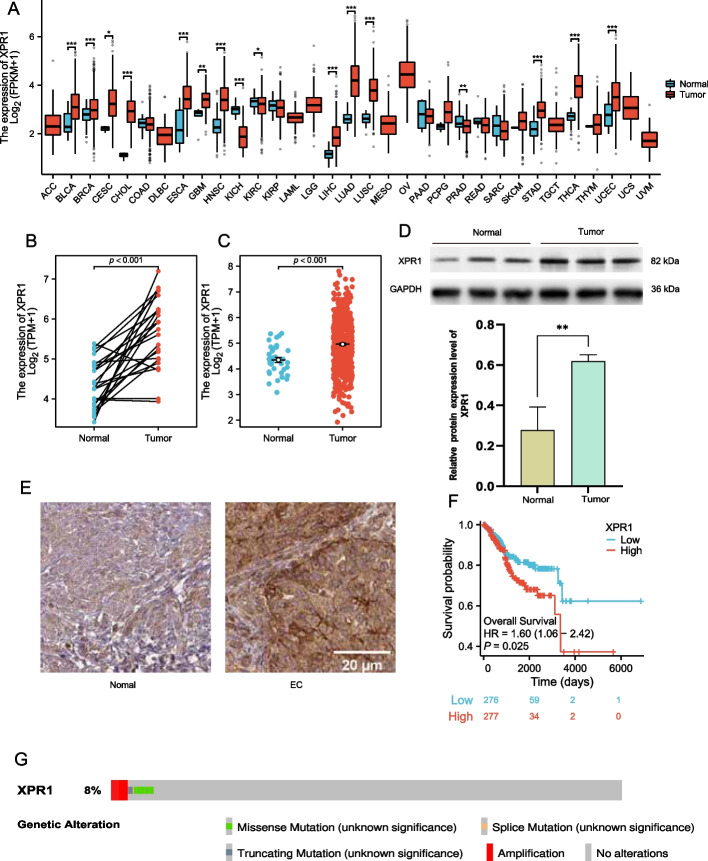
This study investigated XPR1 expression in 554 Uterine Corpus Endometrial Carcinoma(UCEC) cases and 35 normal tissue samples using the The Cancer Genome Atlas(TCGA) database. Western blotting confirmed XPR1 protein presence in EC cell lines (ECC-1) and normal endometrial cells (EEC). The impact of XPR1 on EC cell proliferation and invasion was assessed using EdU proliferation and Transwell invasion assays. A Dot blot assay evaluated m6A methylation of XPR1 in ECC-1 and EEC. The relationship between XPR1 and EC patient prognosis was analyzed using Kaplan-Meier survival and Cox regression analyses. A nomogram was developed to predict 1-, 3-, and 5-year survival probabilities for EC patients, with its accuracy assessed via a calibration curve. Functional enrichment analyses using Gene Ontology (GO) and Kyoto Encyclopedia of Genes and Genomes (KEGG) were conducted to elucidate biological roles and pathways related to XPR1-associated genes. The 'corrplot' R package analyzed the correlation between XPR1 expression and m6A methylation in EC, as well as its link to immune infiltration. Statistical analyses were performed using R software, with a bilateral p-value < 0.05 considered statistically significant.
This study revealed that XPR1 expression was significantly elevated in EC tissues compared to normal tissues (p < 0.001). Its expression correlated with clinical characteristics including patient age, BMI, clinical stage, histological grade, and tumor invasiveness, suggesting its potential as a prognostic marker.Kaplan-Meier analysis demonstrated that high XPR1 expression correlated with reduced overall survival (HR = 1.60, 95% CI: 1.06-2.42, p = 0.025). However, multivariable Cox regression analysis did not identify XPR1 as an independent prognostic factor.Functional experiments indicated that elevated XPR1 expression promoted proliferative and invasive capacities in endometrial carcinoma cells. XPR1 expression also associated with infiltration levels of immune cells (B cells, CD8 + T cells, CD4 + T cells, macrophages, neutrophils, and dendritic cells), suggesting potential involvement in tumor immune microenvironment regulation.Co-expressed genes with XPR1 were enriched in key biological processes including RNA processing, DNA metabolic regulation, and KEGG signaling pathways. Furthermore, XPR1 showed positive correlations with multiple m6A-related genes, and high XPR1 expression in EC coincided with elevated m6A methylation levels. Although these findings indicate that there is a correlation between XPR1 and m6A modification, it is uncertain that XPR1 can directly regulate m6A modification. The specific mechanism of XPR1 in m6A mediated post transcriptional regulation needs further study.
This study identified a significant association between elevated XPR1 expression and adverse clinical outcomes in endometrial carcinoma patients, with additional connections observed to m6A modification and tumor immune microenvironment dynamics. As a novel prognostic indicator, XPR1 demonstrated correlations with tumor aggressiveness features and biological processes-including cellular proliferation, invasion, and m6A/immune-related pathways-suggesting its potential biological relevance in endometrial carcinogenesis. However, its independent prognostic value remains undefined, and direct mechanistic roles in m6A-mediated post-transcriptional modulation warrant subsequent experimental validation.
XPR1在某些肿瘤的发生发展中起关键作用,但其与子宫内膜癌(EC)的关系仍不明确。我们推测XPR1在EC中表达升高,且与患者不良预后显著相关,有望成为一种预后生物标志物。
本研究利用癌症基因组图谱(TCGA)数据库,调查了554例子宫体子宫内膜癌(UCEC)病例和35例正常组织样本中XPR1的表达情况。蛋白质免疫印迹法证实了XPR1蛋白在EC细胞系(ECC-1)和正常子宫内膜细胞(EEC)中的存在。采用EdU增殖实验和Transwell侵袭实验评估XPR1对EC细胞增殖和侵袭的影响。通过斑点印迹法评估ECC-1和EEC中XPR1的m6A甲基化情况。采用Kaplan-Meier生存分析和Cox回归分析来分析XPR1与EC患者预后的关系。绘制列线图以预测EC患者1年、3年和5年的生存概率,并通过校准曲线评估其准确性。利用基因本体论(GO)和京都基因与基因组百科全书(KEGG)进行功能富集分析,以阐明与XPR1相关基因有关的生物学作用和途径。使用“corrplot”R包分析EC中XPR1表达与m6A甲基化之间的相关性,以及其与免疫浸润的关系。使用R软件进行统计分析,双侧p值<0.05被认为具有统计学意义。
本研究表明,与正常组织相比,EC组织中XPR1的表达显著升高(p<0.001)。其表达与患者年龄、BMI、临床分期、组织学分级和肿瘤侵袭性等临床特征相关,表明其具有作为预后标志物的潜力。Kaplan-Meier分析表明,XPR1高表达与总生存期缩短相关(HR=1.60,95%CI:1.06-2.42,p=0.025)。然而,多变量Cox回归分析未将XPR1确定为独立的预后因素。功能实验表明,XPR1表达升高促进了子宫内膜癌细胞的增殖和侵袭能力。XPR1表达还与免疫细胞(B细胞、CD8+T细胞、CD4+T细胞、巨噬细胞、中性粒细胞和树突状细胞)的浸润水平相关,提示其可能参与肿瘤免疫微环境的调节。与XPR1共表达的基因在包括RNA加工、DNA代谢调控等关键生物学过程以及KEGG信号通路中富集。此外,XPR1与多个m6A相关基因呈正相关,且EC中XPR1高表达与m6A甲基化水平升高一致。尽管这些发现表明XPR1与m6A修饰之间存在相关性,但尚不确定XPR1是否能直接调节m6A修饰。XPR1在m6A介导的转录后调控中的具体机制有待进一步研究。
本研究发现XPR1表达升高与子宫内膜癌患者的不良临床结局之间存在显著关联,还观察到其与m6A修饰和肿瘤免疫微环境动态变化有关。作为一种新型预后指标,XPR1与肿瘤侵袭性特征以及包括细胞增殖、侵袭和m6A/免疫相关途径在内的生物学过程相关,提示其在子宫内膜癌发生发展中可能具有生物学相关性。然而,其独立预后价值尚不明确,在m


