Bach Camila A, Hossain Md Niamat, Chaudhari Ishan J, Verrillo Cecilia E, Naranjo Nicole M, Amoroso Isabella, Testa Anna, Sey Samuel, Kelly William K, Bellis Susan L, Lorico Aurelio, Blidner Ada G, Rabinovich Gabriel A, Languino Lucia R
Prostate Cancer Discovery and Development Program, Thomas Jefferson University, Philadelphia, Pennsylvania, United States of America.
Department of Pharmacology, Physiology, and Cancer Biology, Thomas Jefferson University, Philadelphia, Pennsylvania, United States of America.
PLoS One. 2025 Sep 12;20(9):e0329014. doi: 10.1371/journal.pone.0329014. eCollection 2025.
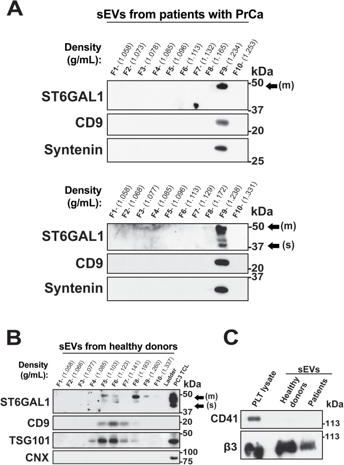
Altered cell surface glycosylation is a hallmark of cancer; among aberrant glycan structures, hypersialylated proteins contribute to disease progression. The enzyme ST6 β-galactoside α2,6-sialyltransferase 1 (ST6GAL1) mediates α2,6-linked sialylation of N-glycosylated proteins and is upregulated in many cancers, including prostate cancer (PrCa). We propose that ST6GAL1 may be released by cancer cells in small extracellular vesicles (sEVs) in the PrCa tumor microenvironment to potentially modulate cell surface sialylation in recipient cells. We isolated sEVs from PrCa cells by density gradient separation and characterized them by nanoparticle tracking analysis using ZetaView and immunoblotting analysis. We identified ST6GAL1 in both its membrane-bound and soluble forms, both active, in circulating sEVs from healthy donors and patients with PrCa. ST6GAL1 is also expressed in human PrCa cells (PC3, DU145, and C4-2B), and in murine cells (TRAMP-C2 and RM1) at different levels, which correlate with aggressive cell phenotypes. In addition to classic sEV markers, such as CD9, TSG101 and Syntenin, sEVs isolated from PrCa cell lines express PDL1, an immune checkpoint ligand. The soluble ST6GAL1 form is present in the sEVs released from DU145 and PC3 cells and can be transferred via sEVs to recipient PrCa cells. This transfer is prevented by expression of Nogo-66 receptor homolog 2 (NgR2) and β3 integrin, which are elevated in the aggressive neuroendocrine phenotype of the disease. The soluble form is absent in the sEVs released from the bone metastatic line C4-2B, which only contains the membrane-bound form. Our results suggest that ST6GAL1 in sEVs derived from PrCa cells may potentially play a role in promoting bone metastasis by facilitating the formation of the pre-metastatic niche.
细胞表面糖基化改变是癌症的一个标志;在异常聚糖结构中,高唾液酸化蛋白促进疾病进展。ST6β-半乳糖苷α2,6-唾液酸转移酶1(ST6GAL1)介导N-糖基化蛋白的α2,6-连接唾液酸化,并且在包括前列腺癌(PrCa)在内的许多癌症中上调。我们提出,ST6GAL1可能由PrCa肿瘤微环境中的癌细胞以小细胞外囊泡(sEVs)的形式释放,从而潜在地调节受体细胞的细胞表面唾液酸化。我们通过密度梯度分离从PrCa细胞中分离出sEVs,并使用ZetaView通过纳米颗粒跟踪分析和免疫印迹分析对其进行表征。我们在来自健康供体和PrCa患者的循环sEVs中鉴定出膜结合形式和可溶性形式的活性ST6GAL1。ST6GAL1也在人PrCa细胞(PC3、DU145和C4-2B)以及鼠细胞(TRAMP-C2和RM1)中以不同水平表达,这与侵袭性细胞表型相关。除了经典的sEV标志物,如CD9、TSG101和Syntenin外,从PrCa细胞系分离的sEVs还表达免疫检查点配体PDL1。可溶性ST6GAL1形式存在于从DU145和PC3细胞释放的sEVs中,并且可以通过sEVs转移到受体PrCa细胞。这种转移被Nogo-66受体同源物2(NgR2)和β3整合素的表达所阻止,它们在该疾病的侵袭性神经内分泌表型中升高。从骨转移细胞系C4-2B释放的sEVs中不存在可溶性形式,其仅包含膜结合形式。我们的结果表明,源自PrCa细胞的sEVs中的ST6GAL1可能通过促进转移前生态位的形成而在促进骨转移中发挥作用。