La Rosa Giuliana, Adorna Miriam, Mauro Letizia Antonella, Pennisi Monica, Musumeci Andrea Giovanni, Sigona Alessandra, Mattina Claudia, Belfiore Giuseppe, Foti Pietro Valerio, Basile Antonio, Palmucci Stefano
Azienda Sanitaria Provinciale di Catania, P.O. "Castiglione-Prestianni", Bronte, Catania, Italy.
Department of Medical Surgical Sciences and Advanced Technologies "GF Ingrassia", Radiology Unit 1, University Hospital Policlinico "G. Rodolico-San Marco", Catania, Italy.
Insights Imaging. 2025 Sep 20;16(1):200. doi: 10.1186/s13244-025-02073-8.
To describe diagnostic and radiological features of the main pathologies affecting the thoracic wall, providing a pictorial atlas based on several clinical cases extracted from our archive.
A wide variety of pathologies affect the tissues of the thoracic cage; these conditions are often encountered by radiologists during examinations performed for unrelated clinical questions. Modern imaging techniques enable the detection of these pathologies and allow definitive diagnoses to be achieved.
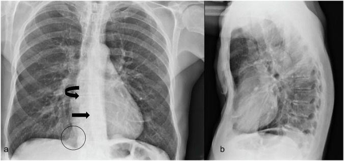
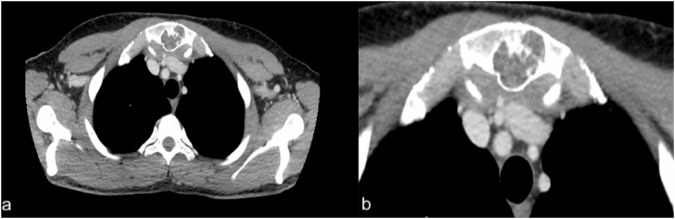
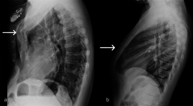
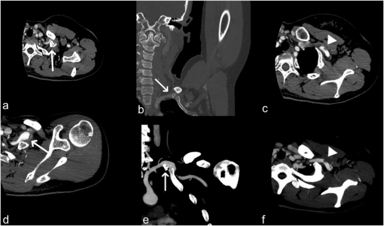
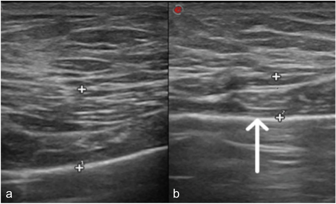
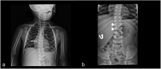
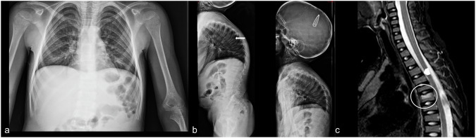
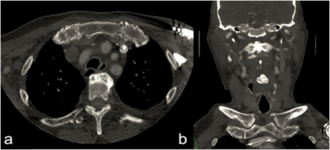
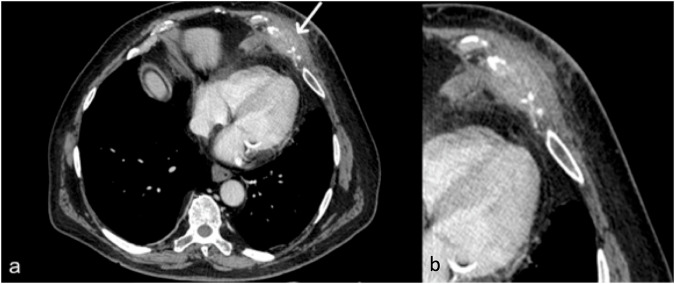
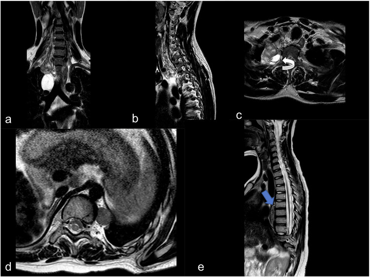
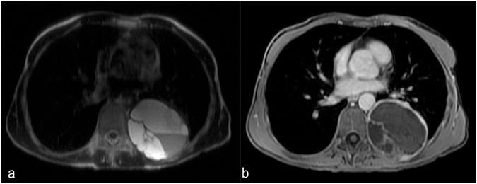
Pathological processes that involve the chest wall may be classified into: (1) congenital and developmental diseases: pectus excavatum, pectus carinatum, supernumerary rib syndrome, Poland syndrome, neurofibromatosis, osteogenesis imperfecta, mucopolysaccharidosis, Marfan syndrome; (2) infectious and inflammatory diseases-such as aspergillosis, tuberculosis, abscesses from pyogenic bacteria, Tietze's syndrome; (3) bone injuries (traumatic and degenerative diseases): sternal, vertebral and costal fractures, degenerative disc and arthrosis pathology; (4) chest wall tumors-such as sarcomas, lymphomas, neurogenic tumors, lipoma.
Thoracic wall pathologies include a wide spectrum of conditions, with some clinical implications that often require a correct nosological framing. Recognizing these pathologies is essential for radiologists so that they can make a correct description in the report and direct toward appropriate treatment if required.
Cage diseases are various and difficult to understand, so multimodality imaging plays a crucial role in achieving an efficient and final diagnosis.
Thoracic wall pathologies have different etiologies. Imaging represents a fundamental tool to clarify their extension, location, and nature. The prognosis of some of these diseases can be poor.
描述影响胸壁的主要病变的诊断和放射学特征,基于从我们的存档中提取的多个临床病例提供一本影像图谱。
多种病变会影响胸廓组织;放射科医生在因不相关临床问题进行的检查中经常会遇到这些情况。现代成像技术能够检测出这些病变并实现明确诊断。
累及胸壁的病理过程可分为:(1)先天性和发育性疾病:漏斗胸、鸡胸、多余肋骨综合征、波兰综合征、神经纤维瘤病、成骨不全、黏多糖贮积症、马方综合征;(2)感染性和炎症性疾病,如曲霉菌病、结核病、化脓性细菌引起的脓肿、蒂策综合征;(3)骨损伤(创伤性和退行性疾病):胸骨、椎体和肋骨骨折、椎间盘退变和关节病病理改变;(4)胸壁肿瘤,如肉瘤、淋巴瘤、神经源性肿瘤、脂肪瘤。
胸壁病变包括广泛的病症,具有一些往往需要正确疾病分类框架的临床意义。对于放射科医生来说,识别这些病变至关重要,以便他们能够在报告中做出正确描述,并在需要时指导进行适当治疗。
胸廓疾病多种多样且难以理解,因此多模态成像在实现高效和最终诊断方面起着至关重要的作用。
胸壁病变有不同的病因。影像学是阐明其范围、位置和性质的基本工具。其中一些疾病的预后可能很差。