• 文献检索
  • 文档翻译
  • 深度研究
  • 学术资讯
  • Suppr Zotero 插件Zotero 插件
  • 邀请有礼
  • 套餐&价格
  • 历史记录
应用&插件
Suppr Zotero 插件Zotero 插件浏览器插件Mac 客户端Windows 客户端微信小程序
定价
高级版会员购买积分包购买API积分包
服务
文献检索文档翻译深度研究API 文档MCP 服务
关于我们
关于 Suppr公司介绍联系我们用户协议隐私条款
关注我们

Suppr 超能文献

核心技术专利:CN118964589B侵权必究
粤ICP备2023148730 号-1Suppr @ 2026

文献检索

告别复杂PubMed语法,用中文像聊天一样搜索,搜遍4000万医学文献。AI智能推荐,让科研检索更轻松。

立即免费搜索

文件翻译

保留排版,准确专业,支持PDF/Word/PPT等文件格式,支持 12+语言互译。

免费翻译文档

深度研究

AI帮你快速写综述,25分钟生成高质量综述,智能提取关键信息,辅助科研写作。

立即免费体验

视网膜电图反应的视网膜下方面

Subretinal aspects of the optoretinographic response.

作者信息

Maddipatla Reddikumar, Bartuzel Maciej M, Pijewska Ewelina, Langlo Christopher S, Zawadzki Robert J, Jonnal Ravi S

出版信息

bioRxiv. 2025 Sep 19:2025.09.18.676889. doi: 10.1101/2025.09.18.676889.

DOI:10.1101/2025.09.18.676889
PMID:41000856
原文链接:https://pmc.ncbi.nlm.nih.gov/articles/PMC12458432/
Abstract

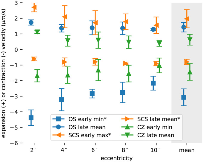
1 Water movement in the living human retina and its regulation are important components of the tissue's structural integrity, optical properties, and homeostasis. In the outer retina there is a continuous flow of water from the subretinal space, through the retinal pigmented epithelium, and into the choroid. This flow is disrupted acutely in disorders such as retinal detachment and central serous retinopathy, and is also known to reduce dramatically with age and age-related macular degeneration. Optoretiongraphy is an emerging technique for measuring neural function in the retina by monitoring nanometer-scale deformations of the membranes of photoreceptors. These deformations have been hypothetically attributed, in part, to osmotic shifts that cause water to move into and out of the photoreceptor outer segment after light stimulation. In the present work, we describe a method for measuring changes in the lengths of the cone outer segment and subretinal space in parallel and results showing that light stimuli change the volume of the subretinal space. These results are consistent with earlier measurements of its light-induced hydration. The magnitude of the latter changes depend on the rate of water clearance from the subretinal space, and thus may serve as an indicator of the health of the water transport system. In addition, they may help us understand the mechanisms underlying the photoreceptor optoretinogram. These findings add to a growing understanding of the ways in which light exposure leads to transient reconfigurations of the outer retinal layers lasting milliseconds to hours.

摘要

1 水在活体人类视网膜中的流动及其调节是该组织结构完整性、光学特性和内环境稳定的重要组成部分。在视网膜外层,存在一股持续的水流,从视网膜下间隙穿过视网膜色素上皮,进入脉络膜。这种流动在视网膜脱离和中心性浆液性视网膜病变等疾病中会急性中断,并且已知随着年龄增长和年龄相关性黄斑变性会显著减少。视网膜电图是一种新兴技术,通过监测光感受器膜的纳米级变形来测量视网膜中的神经功能。这些变形据推测部分归因于渗透变化,这种变化会导致光刺激后水进出光感受器外段。在本研究中,我们描述了一种并行测量视锥细胞外段和视网膜下间隙长度变化的方法,结果表明光刺激会改变视网膜下间隙的体积。这些结果与早期对其光诱导水合作用的测量结果一致。后者变化的幅度取决于视网膜下间隙水清除的速率,因此可能作为水运输系统健康状况的指标。此外,它们可能有助于我们理解光感受器视网膜电图背后的机制。这些发现进一步加深了我们对光暴露导致外层视网膜层持续数毫秒至数小时的短暂重构方式的理解。

https://cdn.ncbi.nlm.nih.gov/pmc/blobs/5e64/12458432/5d2101cea5eb/nihpp-2025.09.18.676889v1-f0006.jpg
https://cdn.ncbi.nlm.nih.gov/pmc/blobs/5e64/12458432/56c13a2c1887/nihpp-2025.09.18.676889v1-f0001.jpg
https://cdn.ncbi.nlm.nih.gov/pmc/blobs/5e64/12458432/7d3134582e7b/nihpp-2025.09.18.676889v1-f0002.jpg
https://cdn.ncbi.nlm.nih.gov/pmc/blobs/5e64/12458432/ad79f5511670/nihpp-2025.09.18.676889v1-f0003.jpg
https://cdn.ncbi.nlm.nih.gov/pmc/blobs/5e64/12458432/b64f58a73cb2/nihpp-2025.09.18.676889v1-f0004.jpg
https://cdn.ncbi.nlm.nih.gov/pmc/blobs/5e64/12458432/129b4d296c35/nihpp-2025.09.18.676889v1-f0005.jpg
https://cdn.ncbi.nlm.nih.gov/pmc/blobs/5e64/12458432/5d2101cea5eb/nihpp-2025.09.18.676889v1-f0006.jpg

相似文献

1
Subretinal aspects of the optoretinographic response.视网膜电图反应的视网膜下方面
bioRxiv. 2025 Sep 19:2025.09.18.676889. doi: 10.1101/2025.09.18.676889.
2
Prescription of Controlled Substances: Benefits and Risks管制药品的处方:益处与风险
3
Vesicoureteral Reflux膀胱输尿管反流
4
Mid Forehead Brow Lift额中眉提升术
5
Shoulder Arthrogram肩关节造影
6
Salzmanns Nodular Corneal Degeneration萨尔茨曼结节状角膜变性
7
Short-Term Memory Impairment短期记忆障碍
8
The Black Book of Psychotropic Dosing and Monitoring.《精神药物剂量与监测黑皮书》
Psychopharmacol Bull. 2024 Jul 8;54(3):8-59.
9
Nonarteritic Anterior Ischemic Optic Neuropathy非动脉炎性前部缺血性视神经病变
10
Aspects of Genetic Diversity, Host Specificity and Public Health Significance of Single-Celled Intestinal Parasites Commonly Observed in Humans and Mostly Referred to as 'Non-Pathogenic'.人类常见且大多被称为“非致病性”的单细胞肠道寄生虫的遗传多样性、宿主特异性及公共卫生意义
APMIS. 2025 Sep;133(9):e70036. doi: 10.1111/apm.70036.

本文引用的文献

1
Velocity-based optoretinography for clinical applications.用于临床应用的基于速度的视网膜电图
Optica. 2022 Oct 20;9(10):1100-1108. doi: 10.1364/optica.460835. Epub 2022 Sep 22.
2
Optical coherence tomography split-spectrum amplitude-decorrelation optoretinography.光学相干断层扫描分光振幅相关光视网膜成像。
Opt Lett. 2023 Aug 1;48(15):3921-3924. doi: 10.1364/OL.492178.
3
Quantification of intrinsic optical signals in the outer human retina using optical coherence tomography.利用光相干断层扫描术对外层人视网膜的固有光学信号进行定量分析。
Ann N Y Acad Sci. 2022 Apr;1510(1):145-157. doi: 10.1111/nyas.14721. Epub 2021 Dec 10.
4
Toward a clinical optoretinogram: a review of noninvasive, optical tests of retinal neural function.迈向临床视网膜电图:视网膜神经功能的非侵入性光学检测综述
Ann Transl Med. 2021 Aug;9(15):1270. doi: 10.21037/atm-20-6440.
5
Functional retinal imaging using adaptive optics swept-source OCT at 1.6 MHz.使用1.6兆赫兹自适应光学扫频源光学相干断层扫描技术进行功能性视网膜成像。
Optica. 2019 Mar 20;6(3):300-303. doi: 10.1364/OPTICA.6.000300.
6
The optoretinogram reveals the primary steps of phototransduction in the living human eye.光视网膜图揭示了活体人眼中光传导的初始步骤。
Sci Adv. 2020 Sep 9;6(37). doi: 10.1126/sciadv.abc1124. Print 2020 Sep.
7
Optoretinogram: optical measurement of human cone and rod photoreceptor responses to light.视激发图:对人眼视锥和视杆光感受器对光反应的光学测量。
Opt Lett. 2020 Sep 1;45(17):4658-4661. doi: 10.1364/OL.398868.
8
The cell biology of the retinal pigment epithelium.视网膜色素上皮细胞生物学
Prog Retin Eye Res. 2020 Feb 24:100846. doi: 10.1016/j.preteyeres.2020.100846.
9
Light-induced changes of the subretinal space of the temporal retina observed via optical coherence tomography.光学相干断层扫描观察到颞侧视网膜光感受器下空间的光诱导变化。
Sci Rep. 2019 Sep 20;9(1):13632. doi: 10.1038/s41598-019-50057-8.
10
In vivo optical imaging of physiological responses to photostimulation in human photoreceptors.人体光感受器对光刺激生理反应的体内光学成像。
Proc Natl Acad Sci U S A. 2016 Nov 15;113(46):13138-13143. doi: 10.1073/pnas.1606428113. Epub 2016 Oct 11.