• 文献检索
  • 文档翻译
  • 深度研究
  • 学术资讯
  • Suppr Zotero 插件Zotero 插件
  • 邀请有礼
  • 套餐&价格
  • 历史记录
应用&插件
Suppr Zotero 插件Zotero 插件浏览器插件Mac 客户端Windows 客户端微信小程序
定价
高级版会员购买积分包购买API积分包
服务
文献检索文档翻译深度研究API 文档MCP 服务
关于我们
关于 Suppr公司介绍联系我们用户协议隐私条款
关注我们

Suppr 超能文献

核心技术专利:CN118964589B侵权必究
粤ICP备2023148730 号-1Suppr @ 2026

文献检索

告别复杂PubMed语法,用中文像聊天一样搜索,搜遍4000万医学文献。AI智能推荐,让科研检索更轻松。

立即免费搜索

文件翻译

保留排版,准确专业,支持PDF/Word/PPT等文件格式,支持 12+语言互译。

免费翻译文档

深度研究

AI帮你快速写综述,25分钟生成高质量综述,智能提取关键信息,辅助科研写作。

立即免费体验

与枯草芽孢杆菌原生质体相关的间体中核糖体的显示。

Demonstration of ribosomes in mesosomes associated with Bacillus subtilis protoplasts.

作者信息

Matheson A, Yang M K, Smith R P

出版信息

J Bacteriol. 1973 Jul;115(1):349-57. doi: 10.1128/jb.115.1.349-357.1973.

DOI:10.1128/jb.115.1.349-357.1973
PMID:4123915
原文链接:https://pmc.ncbi.nlm.nih.gov/articles/PMC246249/
Abstract

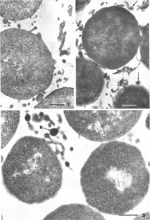
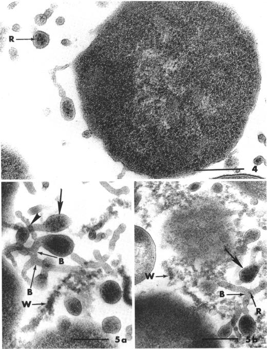
The physiological differences between Bacillus subtilis (ATCC 6633) cells derived from a glucose-salts-yeast extract (GSY) medium and those of cells from tryptose broth permitted the identification of variables in protoplasting environments which noticeably affected the clarity of mesosomal ribosomes. They were the sucrose and magnesium ion concentrations and the type of buffer used. The environment suitable for conversion of GSY cells to the protoplast state was a 0.02 M tris(hydroxymethyl)aminomethane-hydrochloride buffer, pH 7.2, containing 0.6 M sucrose and 0.03 M MgCl(2). Branched mesosomal tubules and a unique organization of vesicles were detected in thin sections and in negative stains of the specimens. Ribosomes were demonstrable in the extruded structures associated with protoplasts that had been prepared according to four fixation schedules and embedded in either of two epoxy plastics. Adjustments in the fixation schedules improved the clarity of the large bodies of protoplast cytoplasm to a degree equivalent to that of their dangling appendages.

摘要

源自葡萄糖 - 盐 - 酵母提取物(GSY)培养基的枯草芽孢杆菌(ATCC 6633)细胞与胰蛋白胨肉汤培养基中的细胞在生理上存在差异,这使得能够确定原生质体形成环境中的变量,这些变量显著影响了中体核糖体的清晰度。这些变量包括蔗糖和镁离子浓度以及所用缓冲液的类型。适合将GSY细胞转化为原生质体状态的环境是含有0.6 M蔗糖和0.03 M MgCl₂的0.02 M三(羟甲基)氨基甲烷 - 盐酸盐缓冲液,pH 7.2。在标本的超薄切片和负染中检测到分支状的中体小管和独特的囊泡组织。在按照四种固定程序制备并包埋在两种环氧塑料中的任何一种中的原生质体相关的挤出结构中,核糖体是可证实的。固定程序的调整将原生质体细胞质大体的清晰度提高到与其悬垂附属物相当的程度。

https://cdn.ncbi.nlm.nih.gov/pmc/blobs/ea6d/246249/9a93d8a89bef/jbacter00347-0366-a.jpg
https://cdn.ncbi.nlm.nih.gov/pmc/blobs/ea6d/246249/a5a7844017d4/jbacter00347-0362-a.jpg
https://cdn.ncbi.nlm.nih.gov/pmc/blobs/ea6d/246249/154b58fc3426/jbacter00347-0363-a.jpg
https://cdn.ncbi.nlm.nih.gov/pmc/blobs/ea6d/246249/a491a026f72e/jbacter00347-0364-a.jpg
https://cdn.ncbi.nlm.nih.gov/pmc/blobs/ea6d/246249/9a93d8a89bef/jbacter00347-0366-a.jpg

相似文献

1
Demonstration of ribosomes in mesosomes associated with Bacillus subtilis protoplasts.与枯草芽孢杆菌原生质体相关的间体中核糖体的显示。
J Bacteriol. 1973 Jul;115(1):349-57. doi: 10.1128/jb.115.1.349-357.1973.
2
Electron-dense particles resembling ribosomes in mesosomes of Bacillus subtilis.在枯草芽孢杆菌中介体中类似核糖体的电子致密颗粒。
J Bacteriol. 1972 May;110(2):747-50. doi: 10.1128/jb.110.2.747-750.1972.
3
Electron microscopy during release and purification of mesosomal vesicles and protoplast membranes from Staphylococcus aureus.金黄色葡萄球菌中体小泡和原生质体膜释放与纯化过程中的电子显微镜观察。
J Bacteriol. 1971 Sep;107(3):907-17. doi: 10.1128/jb.107.3.907-917.1971.
4
ELECTRON MICROSCOPE STUDY OF THE RELATIONSHIP BETWEEN MESOSOME LOSS AND THE STABLE L STATE (OR PROTOPLAST STATE) IN BACILLUS SUBTILIS.枯草芽孢杆菌中质体丢失与稳定L态(或原生质体态)关系的电子显微镜研究
J Bacteriol. 1964 Aug;88(2):457-67. doi: 10.1128/jb.88.2.457-467.1964.
5
Bacteria-shaped gymnoplasts (protoplasts) of Bacillus subtilis.枯草芽孢杆菌的细菌形态裸质体(原生质体)
J Bacteriol. 1969 Jul;99(1):304-15. doi: 10.1128/jb.99.1.304-315.1969.
6
Comparison of the biochemistry and rates of synthesis of mesosomal and peripheral membranes in Bacillus subtilis.枯草芽孢杆菌中介体膜和外周膜的生物化学及合成速率比较。
J Bacteriol. 1971 Jul;107(1):345-57. doi: 10.1128/jb.107.1.345-357.1971.
7
Electron microscopic demonstration and isolation of ribosomes in mesosomes from Staphylococcus aureus.金黄色葡萄球菌中体核糖体的电子显微镜显示及分离
Microbiol Immunol. 1987;31(1):35-44. doi: 10.1111/j.1348-0421.1987.tb03066.x.
8
Electron microscopic studies on protoplast release from I.C.A.-1.65 line (I.F.C.-1.65 line) of Bacillus subtilis cells.
Roum Arch Microbiol Immunol. 1991 Jan-Mar;50(1):27-35.
9
[Isolation conditions for cell nuclei from yeast protoplasts].[从酵母原生质体中分离细胞核的条件]
Z Allg Mikrobiol. 1971;11(2):131-42. doi: 10.1002/jobm.3630110208.
10
Influences of environmental ionic strength, pH, and magnesium ion on bactericidal and lytic activities of -lysin on Bacillus subtilis.环境离子强度、pH值和镁离子对 -溶素对枯草芽孢杆菌的杀菌和裂解活性的影响。
Can J Microbiol. 1972 Nov;18(11):1665-70. doi: 10.1139/m72-258.

引用本文的文献

1
Mesosome structure in Chromobacterium violaceum.紫色色杆菌中的间体结构。
J Bacteriol. 1974 May;118(2):717-24. doi: 10.1128/jb.118.2.717-724.1974.
2
[Electron microscopy of ageing cells of Pseudomonas rhodos: fine structure of native and isolated tubular membranes (author's transl)].
Arch Microbiol. 1975 Aug 28;104(3):215-23. doi: 10.1007/BF00447327.
3
Mesosomes: membranous bacterial organelles.间体:细菌的膜性细胞器。
Bacteriol Rev. 1975 Dec;39(4):405-63. doi: 10.1128/br.39.4.405-463.1975.

本文引用的文献

1
Effect of penicillin on the cell wall of Bacillus subtilis.青霉素对枯草芽孢杆菌细胞壁的作用。
Biochim Biophys Acta. 1962 May 21;59:458-66. doi: 10.1016/0006-3002(62)90196-8.
2
ELECTRON MICROSCOPE STUDY OF THE RELATIONSHIP BETWEEN MESOSOME LOSS AND THE STABLE L STATE (OR PROTOPLAST STATE) IN BACILLUS SUBTILIS.枯草芽孢杆菌中质体丢失与稳定L态(或原生质体态)关系的电子显微镜研究
J Bacteriol. 1964 Aug;88(2):457-67. doi: 10.1128/jb.88.2.457-467.1964.
3
Improvements in epoxy resin embedding methods.环氧树脂包埋方法的改进。
J Biophys Biochem Cytol. 1961 Feb;9(2):409-14. doi: 10.1083/jcb.9.2.409.
4
Electron microscope study of DNA-containing plasms. II. Vegetative and mature phage DNA as compared with normal bacterial nucleoids in different physiological states.含DNA质体的电子显微镜研究。II. 营养期和成熟噬菌体DNA与处于不同生理状态的正常细菌类核的比较。
J Biophys Biochem Cytol. 1958 Nov 25;4(6):671-8. doi: 10.1083/jcb.4.6.671.
5
Morphological phenomena associated with penicillinase induction and secretion in Bacillus licheniformis.与地衣芽孢杆菌中青霉素酶诱导和分泌相关的形态学现象。
J Bacteriol. 1968 Oct;96(4):1314-28. doi: 10.1128/jb.96.4.1314-1328.1968.
6
[Behavior of mesosomes at the time of attack of Bacillus subtilis by lysozyme in a hyper- or hypotonic medium].
C R Acad Hebd Seances Acad Sci D. 1967 Oct 23;265(17):1259-62.
7
Fine structure of Bacillus megaterium during synchronous growth.巨大芽孢杆菌同步生长期间的精细结构
J Bacteriol. 1967 Oct;94(4):1189-205. doi: 10.1128/jb.94.4.1189-1205.1967.
8
Fine structure of Listeria monocytogenes in relation to protoplast formation.与原生质体形成相关的单核细胞增生李斯特菌的精细结构
J Bacteriol. 1967 Jan;93(1):411-26. doi: 10.1128/jb.93.1.411-426.1967.
9
[Morphologic study of the relationship of the membrane wall in E. coli and the protoplasts of B. subtilis].[大肠杆菌细胞膜壁与枯草芽孢杆菌原生质体关系的形态学研究]
Ann Inst Pasteur (Paris). 1966 Jun;110(6):801-12.
10
Some enzymic activities and chemical properties of the mesosomes and cytoplasmic membranes of Bacillus licheniformis 6346.地衣芽孢杆菌6346的间体和细胞质膜的一些酶活性及化学性质
Biochem J. 1969 Jun;113(1):67-79. doi: 10.1042/bj1130067.