• 文献检索
  • 文档翻译
  • 深度研究
  • 学术资讯
  • Suppr Zotero 插件Zotero 插件
  • 邀请有礼
  • 套餐&价格
  • 历史记录
应用&插件
Suppr Zotero 插件Zotero 插件浏览器插件Mac 客户端Windows 客户端微信小程序
定价
高级版会员购买积分包购买API积分包
服务
文献检索文档翻译深度研究API 文档MCP 服务
关于我们
关于 Suppr公司介绍联系我们用户协议隐私条款
关注我们

Suppr 超能文献

核心技术专利:CN118964589B侵权必究
粤ICP备2023148730 号-1Suppr @ 2026

文献检索

告别复杂PubMed语法,用中文像聊天一样搜索,搜遍4000万医学文献。AI智能推荐,让科研检索更轻松。

立即免费搜索

文件翻译

保留排版,准确专业,支持PDF/Word/PPT等文件格式,支持 12+语言互译。

免费翻译文档

深度研究

AI帮你快速写综述,25分钟生成高质量综述,智能提取关键信息,辅助科研写作。

立即免费体验

普氏立克次氏体细胞壁的电子显微镜观察。

Electron microscopy of the cell wall of Rickettsia prowazeki.

作者信息

Palmer E L, Mallavia L P, Tzianabos T, Obijeski J F

出版信息

J Bacteriol. 1974 Jun;118(3):1158-66. doi: 10.1128/jb.118.3.1158-1166.1974.

DOI:10.1128/jb.118.3.1158-1166.1974
PMID:4133612
原文链接:https://pmc.ncbi.nlm.nih.gov/articles/PMC246866/
Abstract

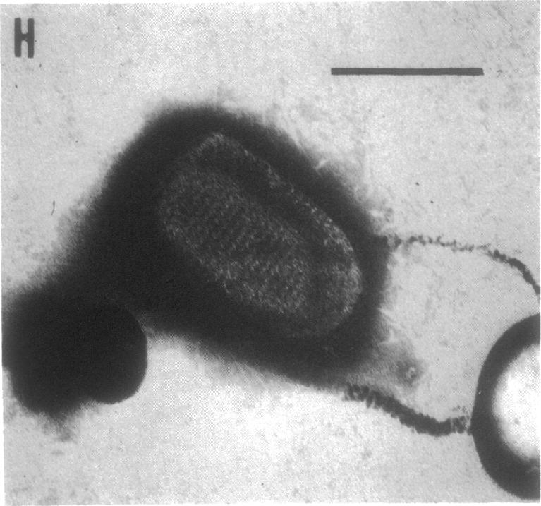
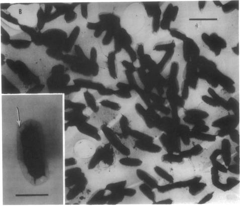
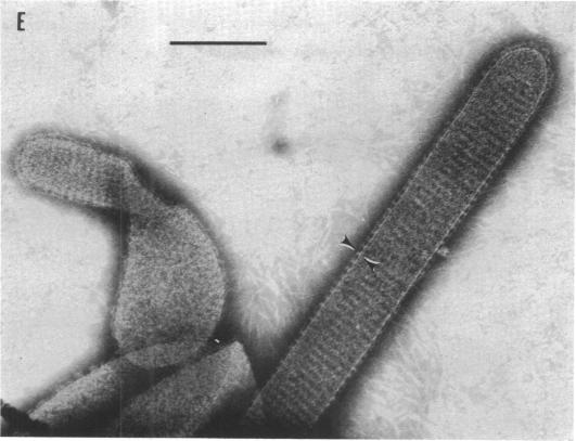
Purified Rickettsia prowazeki were found to undergo morphological changes resembling plasmolysis when stained with uranyl acetate, resulting in rod-like forms. Sequential electron micrographs of disintegrating organisms provide evidence for the cell wall origin of these rod-like forms. The substructure of the cell wall was discerned by using negative-contrast electron microscopy. The wall was found to be composed of repetitive subunits with a periodicity of 13 nm and was surrounded by a thin membrane.

摘要

经发现,用醋酸铀酰染色时,纯化的普氏立克次氏体发生了类似质壁分离的形态变化,形成了杆状形态。对解体生物体的连续电子显微镜照片为这些杆状形态的细胞壁起源提供了证据。通过负染色电子显微镜观察到了细胞壁的亚结构。发现细胞壁由周期为13纳米的重复亚基组成,并被一层薄膜包围。

https://cdn.ncbi.nlm.nih.gov/pmc/blobs/ac91/246866/063f96949d20/jbacter00342-0410-c.jpg
https://cdn.ncbi.nlm.nih.gov/pmc/blobs/ac91/246866/32f8d154f479/jbacter00342-0405-a.jpg
https://cdn.ncbi.nlm.nih.gov/pmc/blobs/ac91/246866/a1c1a0f68286/jbacter00342-0406-a.jpg
https://cdn.ncbi.nlm.nih.gov/pmc/blobs/ac91/246866/2bf26b73ca1d/jbacter00342-0406-b.jpg
https://cdn.ncbi.nlm.nih.gov/pmc/blobs/ac91/246866/cd3882359551/jbacter00342-0407-a.jpg
https://cdn.ncbi.nlm.nih.gov/pmc/blobs/ac91/246866/2580b59bf337/jbacter00342-0408-a.jpg
https://cdn.ncbi.nlm.nih.gov/pmc/blobs/ac91/246866/a1d09b69851a/jbacter00342-0409-a.jpg
https://cdn.ncbi.nlm.nih.gov/pmc/blobs/ac91/246866/067743dabbfd/jbacter00342-0409-b.jpg
https://cdn.ncbi.nlm.nih.gov/pmc/blobs/ac91/246866/14979228d7a4/jbacter00342-0410-a.jpg
https://cdn.ncbi.nlm.nih.gov/pmc/blobs/ac91/246866/2035ba5210ac/jbacter00342-0410-b.jpg
https://cdn.ncbi.nlm.nih.gov/pmc/blobs/ac91/246866/063f96949d20/jbacter00342-0410-c.jpg

相似文献

1
Electron microscopy of the cell wall of Rickettsia prowazeki.普氏立克次氏体细胞壁的电子显微镜观察。
J Bacteriol. 1974 Jun;118(3):1158-66. doi: 10.1128/jb.118.3.1158-1166.1974.
2
Ultrastucture of the surface of Rickettsia prowazeki and Rickettsia akari.普氏立克次体和小蛛立克次体表面的超微结构。
Appl Microbiol. 1974 Oct;28(4):713-6. doi: 10.1128/am.28.4.713-716.1974.
3
New electron microscopic data on structural formations in the cytoplasm of rickettsia prowazeki.关于普氏立克次体细胞质中结构形成的新电子显微镜数据。
Acta Virol. 1971 Nov;15(6):510-4.
4
Electron microscopy of surface structures of Rickettsia prowazeki stained with ruthenium red.
Acta Virol. 1976 Oct;20(5):424-8.
5
Antigens of Rickettsia prowazeki. I. Activity in complement fixation and mouse protection tests.
J Immunol. 1968 Mar;100(3):543-7.
6
Antigens of Rickettsia prowazeki. II. Activity in guinea pigs and exerimental vaccine production.普氏立克次体抗原。II. 在豚鼠体内的活性及实验性疫苗生产
J Immunol. 1968 Mar;100(3):548-53.
7
Fine structure of Rickettsia prowazeki in the haemocytes of ticks Hyalomma dromedarii.热带璃眼蜱血细胞中普氏立克次体的精细结构
Acta Virol. 1967 Jan;11(1):60-2.
8
[Soluble antigens of Rickettsia prowazekii in suspension before and after its treatment with ether].[普氏立克次氏体可溶性抗原在经乙醚处理前后的悬浮液中]
Zh Mikrobiol Epidemiol Immunobiol. 1970 Apr;47(4):110-4.
9
Electron microscopy during release and purification of mesosomal vesicles and protoplast membranes from Staphylococcus aureus.金黄色葡萄球菌中体小泡和原生质体膜释放与纯化过程中的电子显微镜观察。
J Bacteriol. 1971 Sep;107(3):907-17. doi: 10.1128/jb.107.3.907-917.1971.
10
[An electron microscopic study of Rickettsia prowazekii].[普氏立克次体的电子显微镜研究]
Zh Mikrobiol Epidemiol Immunobiol. 1970 Mar;47(3):82-5.

引用本文的文献

1
O antigen is the target of bactericidal Weil-Felix antibodies.O 抗原是杀菌魏尔肖-菲克斯抗体的靶标。
Proc Natl Acad Sci U S A. 2019 Sep 24;116(39):19659-19664. doi: 10.1073/pnas.1911922116. Epub 2019 Aug 14.
2
Comparison of different media for preservation and transport of viable rickettsiae.用于保存和运输活立克次氏体的不同培养基的比较。
Eur J Microbiol Immunol (Bp). 2013 Sep;3(3):194-7. doi: 10.1556/EuJMI.3.2013.3.7. Epub 2013 Sep 23.
3
Inactivation of rickettsiae.立克次氏体的灭活

本文引用的文献

1
Metabolic studies of rickettsiae. I. The effects of antimicrobial substances and enzyme inhibitors on the oxidation of glutamate by purified rickettsiae.立克次氏体的代谢研究。I. 抗菌物质和酶抑制剂对纯化立克次氏体氧化谷氨酸的影响。
J Immunol. 1951 Aug;67(2):123-36.
2
STAINING RICKETTSIAE IN YOLK-SAC CULTURES.卵黄囊培养物中里克次氏体的染色
Stain Technol. 1964 May;39:135-40. doi: 10.3109/10520296409061219.
3
Cell-wall constituents of rickettsiae and psittacosis-lymphogranuloma organisms.立克次氏体和鹦鹉热-淋巴肉芽肿病原体的细胞壁成分。
Eur J Microbiol Immunol (Bp). 2013 Sep;3(3):188-93. doi: 10.1556/EuJMI.3.2013.3.6. Epub 2013 Sep 23.
4
Adherence to and invasion of host cells by spotted Fever group rickettsia species.斑点热群立克次体对宿主细胞的黏附和侵袭
Front Microbiol. 2010 Dec 20;1:139. doi: 10.3389/fmicb.2010.00139. eCollection 2010.
5
Glycosylation of homologous immunodominant proteins of Ehrlichia chaffeensis and Ehrlichia canis.恰菲埃立克体和犬埃立克体同源免疫显性蛋白的糖基化
Infect Immun. 2000 Jan;68(1):13-8. doi: 10.1128/IAI.68.1.13-18.2000.
6
Reproduction and morphogenesis of Rickettsiella chironomi, an unusual intracellular procaryotic parasite of midge larvae.摇蚊立克次氏体(一种摇蚊幼虫不寻常的细胞内原核寄生虫)的繁殖与形态发生
J Bacteriol. 1980 Aug;143(2):995-1002. doi: 10.1128/jb.143.2.995-1002.1980.
7
Isolation of species-specific protein antigens of Rickettsia typhi and Rickettsia prowazekii for immunodiagnosis and immunoprophylaxis.伤寒立克次体和普氏立克次体种特异性蛋白抗原的分离用于免疫诊断和免疫预防。
J Clin Microbiol. 1981 Sep;14(3):333-41. doi: 10.1128/jcm.14.3.333-341.1981.
8
Partial purification and characterization of the major species-specific protein antigens of Rickettsia typhi and Rickettsia prowazekii identified by rocket immunoelectrophoresis.用火箭免疫电泳法鉴定伤寒立克次氏体和普氏立克次氏体主要种特异性蛋白质抗原的部分纯化及特性分析
Infect Immun. 1981 Jan;31(1):276-88. doi: 10.1128/iai.31.1.276-288.1981.
9
Origin and structure of the group-specific, complement-fixing antigen of Rickettsia rickettsii.立氏立克次体群特异性补体结合抗原的起源与结构
Appl Microbiol. 1974 Sep;28(3):481-8. doi: 10.1128/am.28.3.481-488.1974.
10
Ultrastucture of the surface of Rickettsia prowazeki and Rickettsia akari.普氏立克次体和小蛛立克次体表面的超微结构。
Appl Microbiol. 1974 Oct;28(4):713-6. doi: 10.1128/am.28.4.713-716.1974.
J Gen Microbiol. 1963 Mar;30:469-80. doi: 10.1099/00221287-30-3-469.
4
Unstable nucleic acids of Rickettsia mooseri.
Science. 1958 Feb 7;127(3293):282-3. doi: 10.1126/science.127.3293.282.
5
Reversible inactivation of typhus rickettsiae at O C.斑疹伤寒立克次体在0℃时的可逆性灭活
J Bacteriol. 1957 Jan;73(1):56-62. doi: 10.1128/jb.73.1.56-62.1957.
6
Electron microscopically revealed structural elements of Bacterium tularense and their in vitro and in vivo role in immunologic reactions.电子显微镜检查揭示了土拉热弗朗西斯菌的结构成分及其在体外和体内免疫反应中的作用。
J Immunol. 1955 Jul;75(1):7-14.
7
The cell wall of Rickettsia mooseri. I. Morphology and chemical composition.穆氏立克次氏体的细胞壁。I. 形态学与化学成分。
J Bacteriol. 1967 Mar;93(3):1113-8. doi: 10.1128/jb.93.3.1113-1118.1967.
8
Electron microscopy of immune disruption of leptospires: action of complement and lysozyme.钩端螺旋体免疫破坏的电子显微镜观察:补体和溶菌酶的作用
J Bacteriol. 1968 Jun;95(6):2293-309. doi: 10.1128/jb.95.6.2293-2309.1968.
9
Hexagonal pattern in cell walls of Escherichia coli B.大肠杆菌B细胞壁中的六边形图案。
Science. 1967 Jan 27;155(3761):472-4. doi: 10.1126/science.155.3761.472.
10
Studies on the cell wall of Spirillum serpens. 1. Isolation and partial purification of the outermost cell wall layer.对蜿蜒螺菌细胞壁的研究。1. 最外层细胞壁层的分离与部分纯化。
Can J Microbiol. 1970 Oct;16(10):1011-22. doi: 10.1139/m70-171.