• 文献检索
  • 文档翻译
  • 深度研究
  • 学术资讯
  • Suppr Zotero 插件Zotero 插件
  • 邀请有礼
  • 套餐&价格
  • 历史记录
应用&插件
Suppr Zotero 插件Zotero 插件浏览器插件Mac 客户端Windows 客户端微信小程序
定价
高级版会员购买积分包购买API积分包
服务
文献检索文档翻译深度研究API 文档MCP 服务
关于我们
关于 Suppr公司介绍联系我们用户协议隐私条款
关注我们

Suppr 超能文献

核心技术专利:CN118964589B侵权必究
粤ICP备2023148730 号-1Suppr @ 2026

文献检索

告别复杂PubMed语法,用中文像聊天一样搜索,搜遍4000万医学文献。AI智能推荐,让科研检索更轻松。

立即免费搜索

文件翻译

保留排版,准确专业,支持PDF/Word/PPT等文件格式,支持 12+语言互译。

免费翻译文档

深度研究

AI帮你快速写综述,25分钟生成高质量综述,智能提取关键信息,辅助科研写作。

立即免费体验

犬冠状动脉暂时闭塞后的“无复流”现象。

The "no-reflow" phenomenon after temporary coronary occlusion in the dog.

作者信息

Kloner R A, Ganote C E, Jennings R B

出版信息

J Clin Invest. 1974 Dec;54(6):1496-508. doi: 10.1172/JCI107898.

DOI:10.1172/JCI107898
PMID:4140198
原文链接:https://pmc.ncbi.nlm.nih.gov/articles/PMC301706/
Abstract

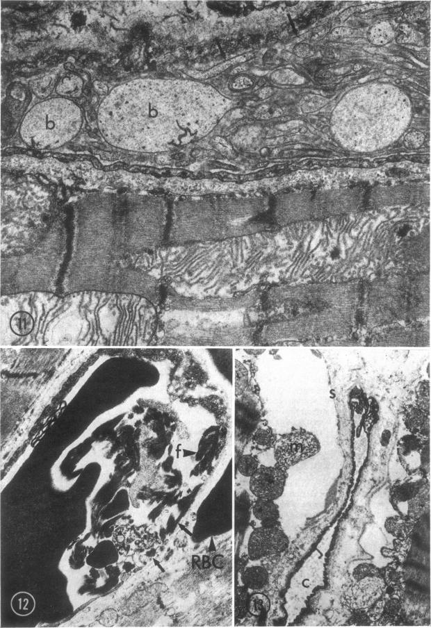
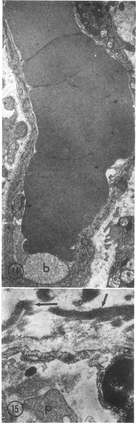
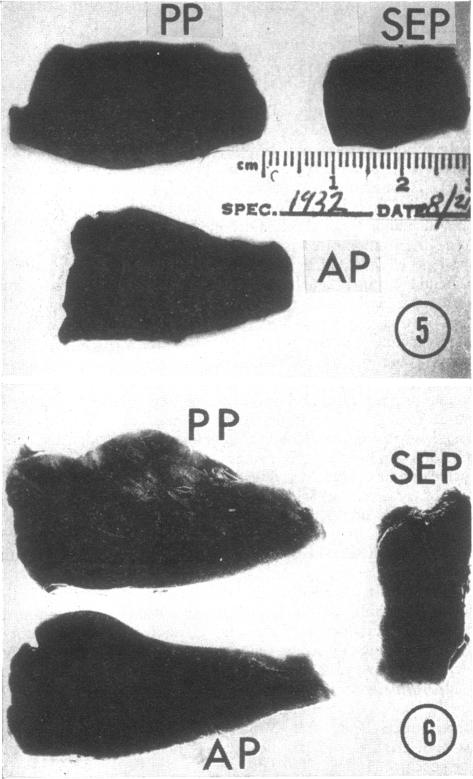
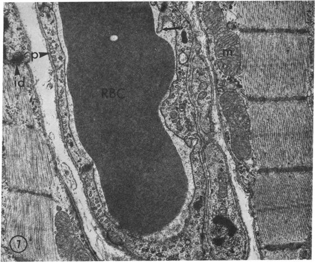
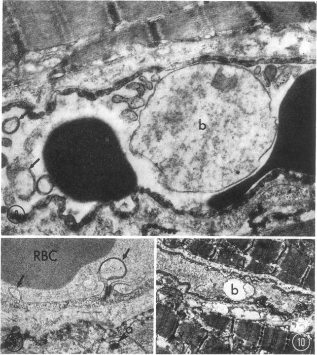
The role of microvascular damage in the genesis of the "no-reflow" phenomenon was investigated in the left ventricular myocardium of dogs subjected to temporary occlusions of a major coronary artery for 40 and 90 min. Intravenous carbon black or thioflavin S (a fluorescent vital stain for endothelium) were used to demonstrate the distribution of coronary arterial flow in control and damaged myocardium. These tracers were injected simultaneously with release of the coronary occlusion or after 5 or 20 min of reflow of coronary arterial blood. After 40 min of ischemia plus arterial reperfusion, usually the tracers were evenly distributed throughout the damaged tissue at each time of reperfusion. On the other hand, when reflow was allowed after 90 min of ischemia, portions of the inner half of damaged myocardium were not penetrated by the tracers. Electron microscopic study of this poorly perfused tissue revealed severe capillary damage; endothelial cells with large intraluminal protrusions and decreased pinocytic vesicles were common. Also, occasional intraluminal fibrin thrombi were noted, as well as extravascular fibrin deposits and erythrocytes. Myocardial cells were swollen in both poorly perfused and well-perfused irreversibly injured tissue. Contraction bands and mitochondrial Ca(2+) accumulation were prominent features of irreversible injury with reflow at 40 min but were not noted after 90 min of ischemia in areas with poor perfusion. These results suggest that 40 min of ischemia were tolerated by the capillary bed of the dog heart without serious capillary damage or perfusion defects, but that 90 min of ischemic injury was associated with the "no-reflow" phenomenon, i.e., failure to achieve uniform reperfusion. This failure of reflow was associated with extensive capillary damage and myocardial cell swelling. Death of severely ischemic myocardial cells in this model occurs before the onset of capillary damage and the no-reflow phenomenon.

摘要

在经历主要冠状动脉临时闭塞40分钟和90分钟的犬左心室心肌中,研究了微血管损伤在“无复流”现象发生中的作用。静脉注射炭黑或硫黄素S(一种内皮细胞荧光活体染料),以显示对照心肌和受损心肌中冠状动脉血流的分布。这些示踪剂在冠状动脉闭塞解除时或冠状动脉血液再灌注5分钟或20分钟后同时注射。缺血40分钟加动脉再灌注后,通常在每次再灌注时示踪剂均匀分布于整个受损组织。另一方面,当缺血90分钟后再灌注时,受损心肌内半部分的一些区域未被示踪剂穿透。对这种灌注不良组织的电子显微镜研究显示严重的毛细血管损伤;内皮细胞内有大的腔内突起且胞饮小泡减少很常见。此外,偶尔可见腔内纤维蛋白血栓以及血管外纤维蛋白沉积和红细胞。在灌注不良和灌注良好的不可逆损伤组织中,心肌细胞均肿胀。收缩带和线粒体Ca(2+)积累是40分钟再灌注时不可逆损伤的突出特征,但在灌注不良区域缺血90分钟后未观察到。这些结果表明,犬心脏的毛细血管床能够耐受40分钟的缺血,而无严重的毛细血管损伤或灌注缺陷,但90分钟的缺血性损伤与“无复流”现象相关,即未能实现均匀再灌注。这种复流失败与广泛的毛细血管损伤和心肌细胞肿胀有关。在该模型中,严重缺血心肌细胞的死亡发生在毛细血管损伤和无复流现象出现之前。

https://cdn.ncbi.nlm.nih.gov/pmc/blobs/a3d4/301706/fe3cbac2d6cb/jcinvest00164-0274-a.jpg
https://cdn.ncbi.nlm.nih.gov/pmc/blobs/a3d4/301706/b324345861d5/jcinvest00164-0270-a.jpg
https://cdn.ncbi.nlm.nih.gov/pmc/blobs/a3d4/301706/fd7cbc763aba/jcinvest00164-0272-a.jpg
https://cdn.ncbi.nlm.nih.gov/pmc/blobs/a3d4/301706/2509f78549aa/jcinvest00164-0265-a.jpg
https://cdn.ncbi.nlm.nih.gov/pmc/blobs/a3d4/301706/3ee1c08a405b/jcinvest00164-0266-a.jpg
https://cdn.ncbi.nlm.nih.gov/pmc/blobs/a3d4/301706/231c11c62e82/jcinvest00164-0267-a.jpg
https://cdn.ncbi.nlm.nih.gov/pmc/blobs/a3d4/301706/f56f26caf381/jcinvest00164-0268-a.jpg
https://cdn.ncbi.nlm.nih.gov/pmc/blobs/a3d4/301706/fe3cbac2d6cb/jcinvest00164-0274-a.jpg

相似文献

1
The "no-reflow" phenomenon after temporary coronary occlusion in the dog.犬冠状动脉暂时闭塞后的“无复流”现象。
J Clin Invest. 1974 Dec;54(6):1496-508. doi: 10.1172/JCI107898.
2
Demonstration of the "no-reflow" phenomenon in the dog heart after temporary ischemia.犬心脏短暂缺血后“无复流”现象的演示。
Recent Adv Stud Cardiac Struct Metab. 1975;10:463-74.
3
Progressive impairment of regional myocardial perfusion after initial restoration of postischemic blood flow.缺血后血流最初恢复后,局部心肌灌注的进行性损害。
Circulation. 1989 Dec;80(6):1846-61. doi: 10.1161/01.cir.80.6.1846.
4
Role of leukocytes in response to acute myocardial ischemia and reflow in dogs.白细胞在犬急性心肌缺血及再灌注反应中的作用
Am J Physiol. 1986 Aug;251(2 Pt 2):H314-23. doi: 10.1152/ajpheart.1986.251.2.H314.
5
Leukocyte capillary plugging in myocardial ischemia and reperfusion in the dog.犬心肌缺血再灌注时的白细胞毛细血管阻塞
Am J Pathol. 1983 Apr;111(1):98-111.
6
Effect of a transient period of ischemia on myocardial cells. II. Fine structure during the first few minutes of reflow.短暂缺血期对心肌细胞的影响。II. 再灌注最初几分钟内的精细结构
Am J Pathol. 1974 Mar;74(3):399-422.
7
[Coronary microcirculation in myocardial reperfusion. The phenomenon of no-reperfusion].[心肌再灌注中的冠状动脉微循环。无再灌注现象]
Arq Bras Cardiol. 1991 Jun;56(6):471-6.
8
Early membrane damage during coronary reperfusion in dogs. Detection by radiolabeled anticardiac myosin (Fab')2.犬冠状动脉再灌注期间的早期膜损伤。通过放射性标记的抗心肌肌球蛋白(Fab')2检测。
J Clin Invest. 1983 Aug;72(2):535-44. doi: 10.1172/JCI111001.
9
Distribution of coronary arterial flow in acute myocardial ischemia.急性心肌缺血时冠状动脉血流的分布
Arch Pathol. 1975 Feb;99(2):86-94.
10
The no reflow phenomenon: a time-limiting factor for reperfusion after coronary occlusion?无复流现象:冠状动脉闭塞后再灌注的时间限制因素?
Am J Cardiol. 1980 Nov;46(5):800-6. doi: 10.1016/0002-9149(80)90431-2.

引用本文的文献

1
Advances in Pathophysiology and Novel Therapeutic Strategies for Coronary No-Reflow Phenomenon.冠状动脉无复流现象的病理生理学进展及新型治疗策略
Biomedicines. 2025 Jul 14;13(7):1716. doi: 10.3390/biomedicines13071716.
2
The contribution of cardiomyocyte hypercontracture to the burden of acute myocardial infarction: an update.心肌细胞过度收缩对急性心肌梗死负担的影响:最新进展
Basic Res Cardiol. 2025 Jun 7. doi: 10.1007/s00395-025-01120-1.
3
Cardioprotection by poloxamer 188 is mediated through increased endothelial nitric oxide production.

本文引用的文献

1
Cytologic studies with the phase Microscope; the formation of blisters on cells in suspension, photocytosis, with observations on the nature of the cellular membrane.用相差显微镜进行的细胞学研究;悬浮细胞上水泡的形成、光细胞作用以及对细胞膜性质的观察。
Am J Pathol. 1948 May;24(3):545-67.
2
Demonstration of blood vessels and lymphatics with a fluorescent dye in ultraviolet light.在紫外线下用荧光染料显示血管和淋巴管。
Anat Rec. 1949 Nov;105(3):433-43, incl 2 pl. doi: 10.1002/ar.1091050305.
3
A COMPARATIVE STUDY OF THE FINE STRUCTURE OF NORMAL AND ISCHEMIC DOG MYOCARDIUM WITH SPECIAL REFERENCE TO EARLY CHANGES FOLLOWING TEMPORARY OCCLUSION OF A CORONARY ARTERY.
泊洛沙姆188的心脏保护作用是通过增加内皮型一氧化氮的生成来介导的。
Sci Rep. 2025 Apr 30;15(1):15170. doi: 10.1038/s41598-025-97079-z.
4
Prediction of myocardial ischemia-reperfusion injury post-PCI: role of sST2 levels in STEMI patients.经皮冠状动脉介入治疗后心肌缺血再灌注损伤的预测:可溶性ST2水平在ST段抬高型心肌梗死患者中的作用
BMC Cardiovasc Disord. 2025 Apr 11;25(1):280. doi: 10.1186/s12872-025-04708-6.
5
Impact of size-dependent differences in excimer laser coronary angioplasty in ST-elevation acute myocardial infarction: nuclear scintigraphy findings.准分子激光冠状动脉成形术中大小依赖性差异对ST段抬高型急性心肌梗死的影响:核素闪烁显像结果
Postepy Kardiol Interwencyjnej. 2025 Mar;21(1):45-54. doi: 10.5114/aic.2025.147979. Epub 2025 Feb 28.
6
Reperfused Myocardial Infarction: The Road to CCS Classification of Acute MI and Beyond.再灌注性心肌梗死:急性心肌梗死及其他情况的CCS分类之路。
JACC Adv. 2025 Feb;4(2):101528. doi: 10.1016/j.jacadv.2024.101528.
7
Molecular Insights into Ischemia-Reperfusion Injury in Coronary Artery Disease: Mechanisms and Therapeutic Implications: A Comprehensive Review.冠状动脉疾病缺血再灌注损伤的分子见解:机制与治疗意义:综述
Antioxidants (Basel). 2025 Feb 13;14(2):213. doi: 10.3390/antiox14020213.
8
IVUS derived plaque characteristics and outcomes in patients with acute coronary syndrome undergoing percutaneous intervention.血管内超声衍生的斑块特征及接受经皮介入治疗的急性冠状动脉综合征患者的预后
Indian Heart J. 2025 Jan-Feb;77(1):36-41. doi: 10.1016/j.ihj.2025.02.001. Epub 2025 Feb 4.
9
Causes of long-term mortality in patients with ST-segment elevation myocardial infarction is dictated by the presence of microvascular obstruction.ST段抬高型心肌梗死患者的长期死亡原因取决于微血管阻塞的存在。
Eur Heart J Open. 2025 Jan 23;5(1):oeaf002. doi: 10.1093/ehjopen/oeaf002. eCollection 2025 Jan.
10
Antiedemic Effect of the Myosin Light Chain Kinase Inhibitor PIK7 in the Rat Model of Myocardial Ischemia Reperfusion Injury.肌球蛋白轻链激酶抑制剂PIK7在大鼠心肌缺血再灌注损伤模型中的抗水肿作用
Curr Issues Mol Biol. 2025 Jan 6;47(1):33. doi: 10.3390/cimb47010033.
正常与缺血犬心肌超微结构的比较研究——特别参考冠状动脉暂时闭塞后的早期变化
Am J Pathol. 1965 Mar;46(3):367-86.
4
FINE STRUCTURAL CHANGES IN MYOCARDIAL ISCHEMIC INJURY.心肌缺血性损伤的超微结构变化
Arch Pathol. 1965 Feb;79:135-43.
5
Myocardial ischemia and early infarction: an electron microscopic study.心肌缺血与早期梗死:一项电子显微镜研究
Am J Pathol. 1959 May-Jun;35(3):489-523.
6
The cytotoxic action of immune gamma globulin and complement on Krebs ascites tumor cells. I. Ultrastructural studies.免疫γ球蛋白和补体对克雷布斯腹水肿瘤细胞的细胞毒性作用。I. 超微结构研究。
J Exp Med. 1959 May 1;109(5):505-10. doi: 10.1084/jem.109.5.505.
7
Blood supply of the myocardium after temporary coronary occlusion.冠状动脉暂时闭塞后心肌的血液供应。
Circ Res. 1966 Jul;19(1):57-62. doi: 10.1161/01.res.19.1.57.
8
Ischemia of the skin. Electron microscopic study of vascular injury.皮肤缺血。血管损伤的电子显微镜研究。
Am J Pathol. 1969 Mar;54(3):327-53.
9
Cerebral ischemia. I. An improved experimental method for study; cardiovascular effects and demonstration of an early vascular lesion in the rabbit.脑缺血。I. 一种改进的研究实验方法;对家兔心血管的影响及早期血管病变的证明
J Neurosurg. 1968 Feb;28(2):150-7. doi: 10.3171/jns.1968.28.2.0150.
10
Cerebral ischemia. III. Vascular changes.脑缺血。III. 血管变化。
Am J Pathol. 1968 Feb;52(2):455-76.