Matilla A, Molland E A
J Clin Pathol. 1974 Sep;27(9):698-709. doi: 10.1136/jcp.27.9.698.
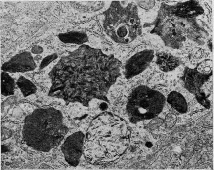
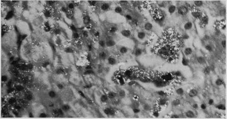
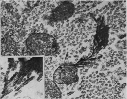
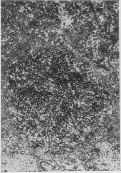
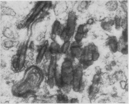
The light- and electron-microscopic appearances of a liver biopsy from a case of erythrohepatic protoporphyria are compared with the appearances of the liver in griseofulvin-induced porphyria in mice. Crystals are present in hepatocytes and in the bile secretory pathways in each case. The nature and significance of these is discussed.
将一例红细胞生成性原卟啉症患者肝脏活检的光镜和电镜表现与灰黄霉素诱导的小鼠卟啉症肝脏表现进行了比较。在每种情况下,肝细胞和胆汁分泌途径中均存在晶体。讨论了这些晶体的性质和意义。