Carpentier J L, Gorden P, Freychet P, Le Cam A, Orci L
J Clin Invest. 1979 Jun;63(6):1249-61. doi: 10.1172/JCI109420.
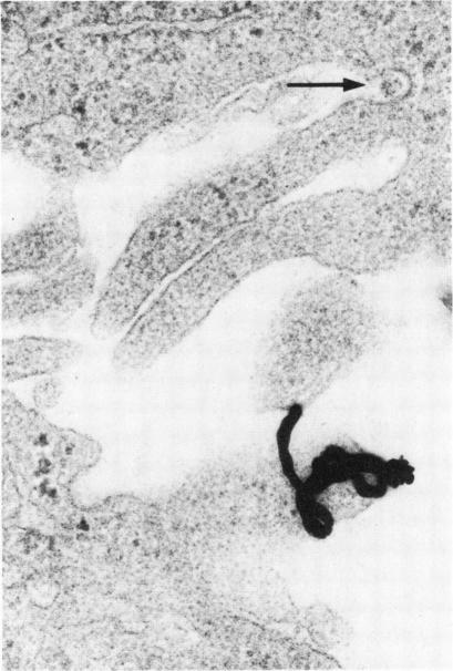
Quantitative electron microscopic autoradiographic studies in cultured human lymphocytes and isolated rat hepatocytes have demonstrated that labeled insulin initially localizes to the plasma membrane and is subsequently internalized to a limited region of the peripheral cytoplasm. When 0.5 nm 125I-insulin is incubated with isolated rat hepatocytes, binding to the plasma membrane occurs at both 20 degrees C and 37 degrees C. Under steady-state binding conditions approximately equal to 30--40% of the labeled hormone is internalized to a distance of approximately equal to 15% of the radius of the cell. When the localization of the internalized labeled material is analyzed, by 2--5 min of incubation at 37 degrees C there is a fivefold preferential association of autoradiographic grains with lysosomal structures, and by 30--60 min of incubation at 37 degrees C there is a 10-fold preferential association. When the cell-associated radioactivity is extracted and filtered on Sephadex G-50 at each time point of incubation, radioactivity elutes predominantly in the position of 125I-insulin and is predominantly in the position of 125I-insulin and is predominantly trichloracetic acid precipitable, bindable to talc, and rebindable to liver membranes. With increasing time of association at 37 degrees C the initial rate and absolute amount of labeled material dissociable from the cell is reduced. With increasing time of dissociation both the cell-associated radioactivity and the radioactivity released into the incubation medium is progressively degraded. These data demonstrate that in isolated rat hepatocytes labeled insulin initially localizes to the plasma membrane, is progressively internalized, and associates preferentially with lysosomal structures. These events may provide a mechanism that links cell surface binding to the degradation of insulin and to insulin-induced loss of its specific receptor.
对培养的人淋巴细胞和分离的大鼠肝细胞进行的定量电子显微镜放射自显影研究表明,标记的胰岛素最初定位于质膜,随后内化到外周细胞质的有限区域。当将0.5纳米的125I-胰岛素与分离的大鼠肝细胞一起孵育时,在20℃和37℃下均会发生与质膜的结合。在稳态结合条件下,约30%-40%的标记激素内化到距细胞半径约15%的距离处。当分析内化标记物质的定位时,在37℃孵育2-5分钟时,放射自显影颗粒与溶酶体结构的优先结合增加了五倍,而在37℃孵育30-60分钟时,优先结合增加了10倍。在孵育的每个时间点提取细胞相关放射性并在Sephadex G-50上过滤时,放射性主要在125I-胰岛素的位置洗脱,并且主要是三氯乙酸可沉淀的,可与滑石粉结合,并可重新结合到肝细胞膜上。随着在37℃下结合时间的增加,可从细胞中解离的标记物质的初始速率和绝对量降低。随着解离时间的增加,细胞相关放射性和释放到孵育培养基中的放射性均逐渐降解。这些数据表明,在分离的大鼠肝细胞中,标记的胰岛素最初定位于质膜,逐渐内化,并优先与溶酶体结构结合。这些事件可能提供了一种机制,将细胞表面结合与胰岛素的降解以及胰岛素诱导的其特异性受体的丧失联系起来。