Hitchins A D, Kahn A J, Slepecky R A
J Bacteriol. 1968 Nov;96(5):1811-7. doi: 10.1128/jb.96.5.1811-1817.1968.
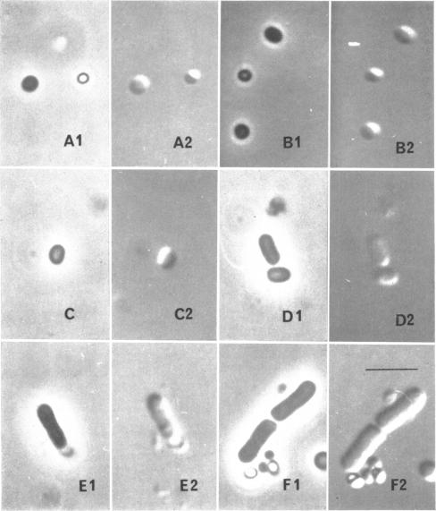
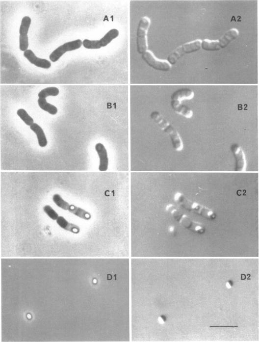
The techniques of Nomarski interference contrast microscopy and phase-contrast microscopy were compared for their utility in monitoring sporulation and germination in Bacillus megaterium. The Nomarski technique permitted rapid and easy delineation of septation and engulfment during sporulation, whereas with phase contrast microscopy these stages were not detected at all. The later stages of sporulation were easily seen by either technique. Thus, of the seven stages of sporulation as recognized by the electron microscopy of thin sections, five can now be routinely detected quantitatively by optical microscopy: septation (stage II), engulfment (stage III), phase-dark forespore (corresponding to cortex formation, stage IV), phase-bright spore in a sporangium (corresponding to coat formation, stage V), and the free spore (stage VII). This means that now only stage I (axial filament) and stage VI (maturation of the refractile spore) require electron microscopy for routine detection. There was no advantage in using Nomarski optics for germination studies.
比较了诺马斯基干涉对比显微镜和相差显微镜技术在监测巨大芽孢杆菌孢子形成和萌发方面的效用。诺马斯基技术能够快速、轻松地描绘出孢子形成过程中的隔膜形成和吞噬现象,而在相差显微镜下根本检测不到这些阶段。孢子形成的后期阶段用这两种技术都很容易观察到。因此,在通过薄切片电子显微镜识别出的孢子形成的七个阶段中,现在有五个阶段可以通过光学显微镜进行常规定量检测:隔膜形成(第二阶段)、吞噬(第三阶段)、暗相前芽孢(对应于皮层形成,第四阶段)、孢子囊内的亮相孢子(对应于孢衣形成,第五阶段)和游离孢子(第七阶段)。这意味着现在只有第一阶段(轴向丝)和第六阶段(折射性孢子的成熟)需要通过电子显微镜进行常规检测。在萌发研究中使用诺马斯基光学显微镜没有优势。