Tibbetts C, Giam C Z
J Virol. 1979 Dec;32(3):995-1005. doi: 10.1128/JVI.32.3.995-1005.1979.
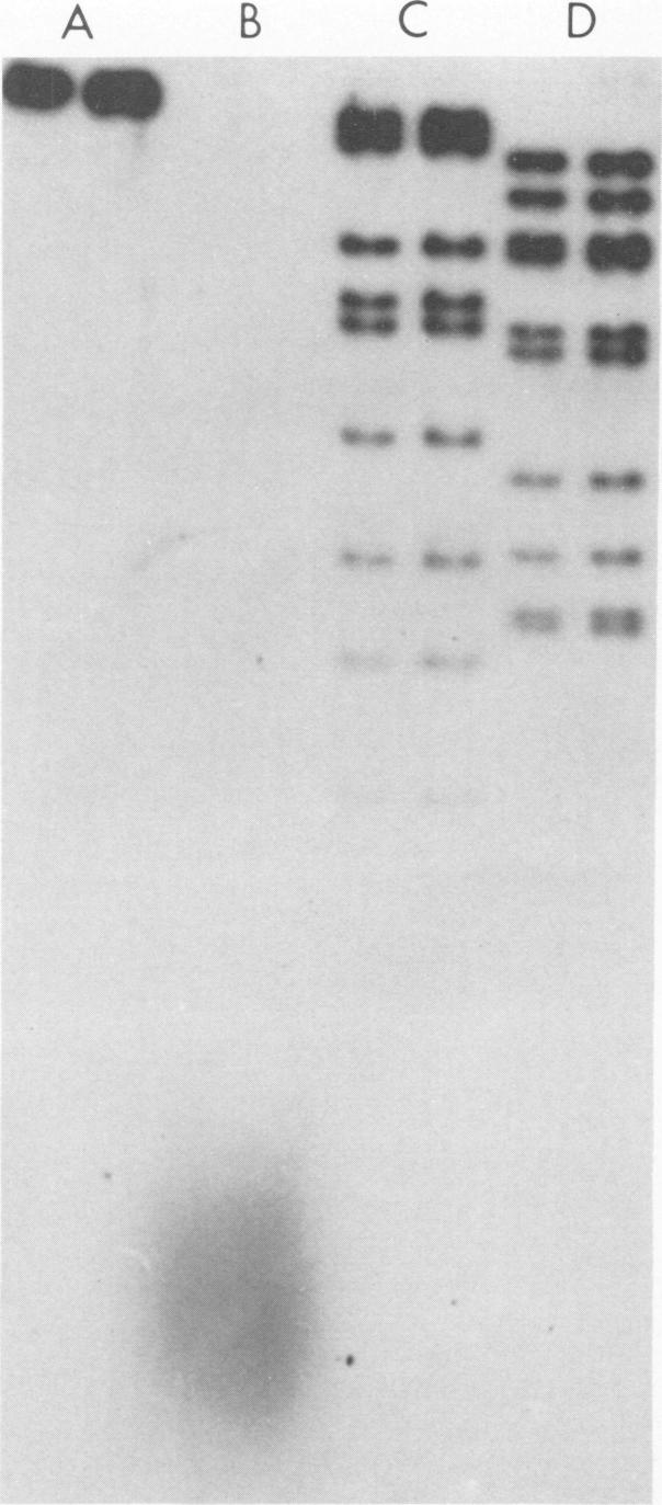
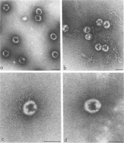
Several lines of evidence suggest that empty adenovirus capsids are preassembled intermediates in the pathway of virion assembly. We have observed that purified empty capsids of subgroup B adenoviruses have a remarkable affinity for DNA in vitro. The products of capsid-DNA association are sufficiently stable, once formed in low-salt solution, to permit purification and characterization in CsCl density gradients. Neither virions nor the DNA-containing incomplete particles of subgroup B adenoviruses can give rise to such in vitro reaction products. The average molecular weight of the empty adenovirus capsids is about 123 X 10(6), consistent with the absence of viral core peptides and a small deficiency of exterior shell polypeptides. Electron microscopy of negatively stained capsids and the capsids bound to DNA reveals a typical adenovirus size and architecture. The particles appear with a surface discontinuity that is presumed to expose the DNA binding site(s). The DNA molecules associated with the empty capsids are susceptible to the actions of DNase and restriction endonucleases. The dependence of rate of capsid-DNA association on DNA length suggests randomly distributed binding sites on the DNA molecules. Although the DNA molecules can successively acquire additional empty capsids, the empty particles themselves are restricted to interactionwith only one DNA molecule. Electron microscopy of the capsid-DNA complexes spread in cytochrome c films shows that the particles are bo-nd along the contour of extended duplex DNA. The amount of DNA within each bound particle appears to be less than 300 base pairs, as estimated by the length of the DNA molecules visible outside of the bound particle. The empty capsid-DNA association product described in this report provides an interesting substrate for further investigation of the DNA packaging process in a defined in vitro system, with extracts or purified components from infected cells.
多条证据表明,空腺病毒衣壳是病毒体组装途径中的预组装中间体。我们观察到,纯化的B亚组腺病毒空衣壳在体外对DNA具有显著的亲和力。衣壳与DNA结合的产物一旦在低盐溶液中形成,就足够稳定,能够在CsCl密度梯度中进行纯化和表征。B亚组腺病毒的病毒体和含DNA的不完全颗粒都不会产生这种体外反应产物。空腺病毒衣壳的平均分子量约为123×10⁶,这与缺乏病毒核心肽以及外壳多肽略有不足一致。对负染衣壳和与DNA结合的衣壳进行电子显微镜观察,揭示了典型的腺病毒大小和结构。这些颗粒出现表面不连续,推测该不连续处暴露了DNA结合位点。与空衣壳相关的DNA分子易受DNA酶和限制性内切酶的作用。衣壳与DNA结合速率对DNA长度的依赖性表明DNA分子上的结合位点是随机分布的。尽管DNA分子可以相继获得额外的空衣壳,但空颗粒本身仅限于与一个DNA分子相互作用。在细胞色素c膜中铺展的衣壳-DNA复合物的电子显微镜观察表明,颗粒沿着延伸的双链DNA轮廓结合。根据结合颗粒外部可见的DNA分子长度估计,每个结合颗粒内的DNA量似乎小于300个碱基对。本报告中描述的空衣壳-DNA结合产物为在体外特定系统中,利用感染细胞的提取物或纯化成分进一步研究DNA包装过程提供了一个有趣的底物。