Tan K B
J Virol. 1970 May;5(5):632-8. doi: 10.1128/JVI.5.5.632-638.1970.
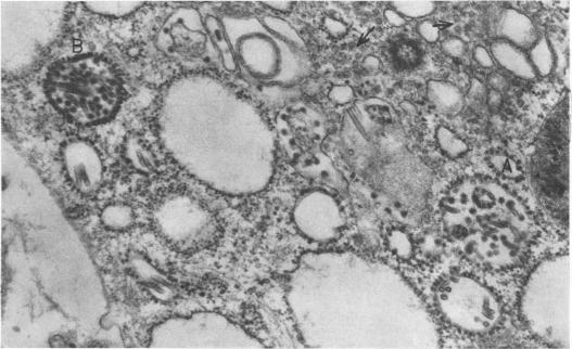
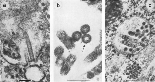
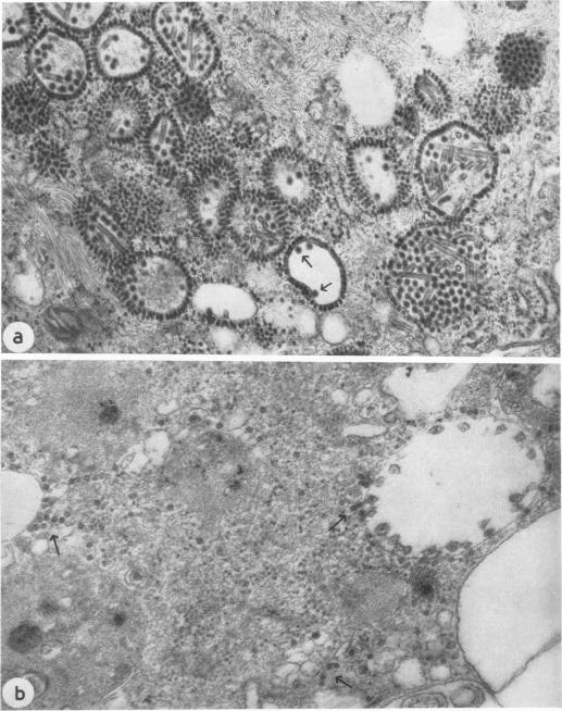
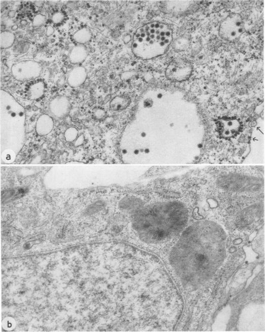
Cells infected at the permissive temperature with three temperature-sensitive mutants of Semliki Forest virus were not significantly different in appearance from cells infected, at either the permissive or nonpermissive temperature, with wild-type virus. Virus particles, nucleocapsids, spherules, and tubules were seen in the cytoplasm. But replication of the mutants was inhibited in cells infected at the nonpermissive temperature. This was evidenced by the absence of virus particles and nucleocapsids (except in one case) and the absence or limited production of spherules and tubules. These observations are discussed with reference to the physiological defects of the mutants.
在允许温度下用三种辛德毕斯病毒温度敏感突变体感染的细胞,在外观上与在允许温度或非允许温度下用野生型病毒感染的细胞没有显著差异。在细胞质中可见病毒颗粒、核衣壳、小球体和小管。但在非允许温度下感染的细胞中,突变体的复制受到抑制。这表现为没有病毒颗粒和核衣壳(只有一例除外),以及小球体和小管的缺失或产生受限。结合突变体的生理缺陷对这些观察结果进行了讨论。