• 文献检索
  • 文档翻译
  • 深度研究
  • 学术资讯
  • Suppr Zotero 插件Zotero 插件
  • 邀请有礼
  • 套餐&价格
  • 历史记录
应用&插件
Suppr Zotero 插件Zotero 插件浏览器插件Mac 客户端Windows 客户端微信小程序
定价
高级版会员购买积分包购买API积分包
服务
文献检索文档翻译深度研究API 文档MCP 服务
关于我们
关于 Suppr公司介绍联系我们用户协议隐私条款
关注我们

Suppr 超能文献

核心技术专利:CN118964589B侵权必究
粤ICP备2023148730 号-1Suppr @ 2026

文献检索

告别复杂PubMed语法,用中文像聊天一样搜索,搜遍4000万医学文献。AI智能推荐,让科研检索更轻松。

立即免费搜索

文件翻译

保留排版,准确专业,支持PDF/Word/PPT等文件格式,支持 12+语言互译。

免费翻译文档

深度研究

AI帮你快速写综述,25分钟生成高质量综述,智能提取关键信息,辅助科研写作。

立即免费体验

细菌L型及其亲本细菌的结构。

Structure of bacterial L forms and their parent bacteria.

作者信息

Weibull C

出版信息

J Bacteriol. 1965 Nov;90(5):1467-80. doi: 10.1128/jb.90.5.1467-1480.1965.

DOI:10.1128/jb.90.5.1467-1480.1965
PMID:5848335
原文链接:https://pmc.ncbi.nlm.nih.gov/articles/PMC315836/
Abstract

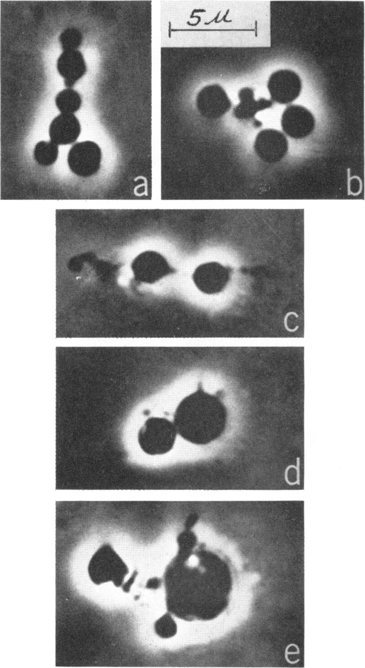
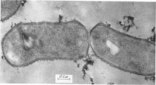
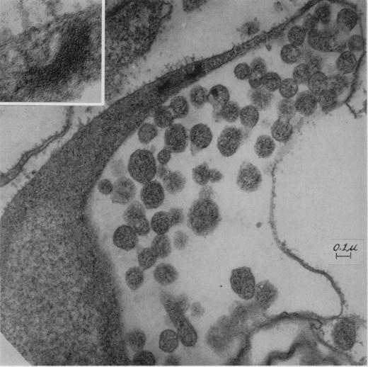
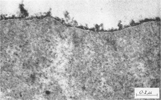
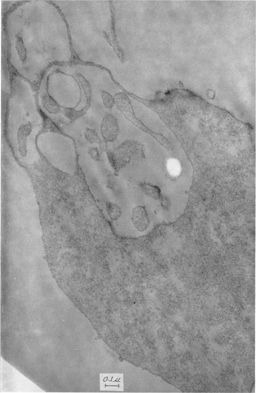
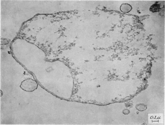
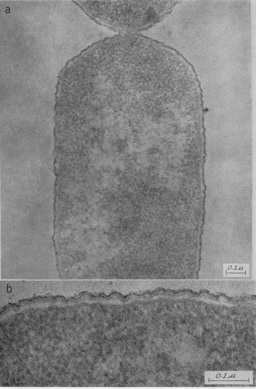
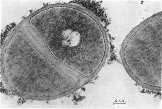
Weibull, Claes (Rocky Mountain Laboratory, Hamilton, Mont.). Structure of bacterial L forms and their parent bacteria. J. Bacteriol. 90:1467-1480. 1965.-Light and electron microscopic studies were done on normal cells and L forms of Proteus mirabilis, Staphylococcus aureus, and Corynebacterium sp. grown in liquid media. Under the prevailing growth conditions, the L forms studied were morphologically indistinguishable from one another. They appeared as approximately spherical elements occurring singly or more often connected with each other by thinner portions of cell material. In sections of large L forms, the following structures were seen: a peripheral, triple-layered ("unit") membrane, a granular cytoplasm, nuclear regions, and vacuoles limited by membranes. Small bodies often were present inside the vacuoles. These bodies also contained a peripheral membrane and a granular cytoplasm but usually no nuclear regions. The normal bacteria from which the L forms were derived differed markedly in structure from one another, especially in the surface layers of the cells.

摘要

魏布尔,克莱斯(落基山实验室,蒙大拿州汉密尔顿)。细菌L型及其亲本细菌的结构。《细菌学杂志》90:1467 - 1480。1965年。- 对奇异变形杆菌、金黄色葡萄球菌和棒状杆菌属在液体培养基中生长的正常细胞和L型进行了光学显微镜和电子显微镜研究。在当时的生长条件下,所研究的L型在形态上彼此难以区分。它们呈现为近似球形的个体,单个出现或更常见的是通过较细的细胞物质部分相互连接。在大型L型的切片中,可见以下结构:外周的三层(“单位”)膜、颗粒状细胞质、核区以及由膜界定的液泡。液泡内常存在小体。这些小体也含有外周膜和颗粒状细胞质,但通常没有核区。L型所源自的正常细菌在结构上彼此差异显著,尤其是在细胞的表层。

https://cdn.ncbi.nlm.nih.gov/pmc/blobs/88d1/315836/56ed3ed86484/jbacter00428-0354-a.jpg
https://cdn.ncbi.nlm.nih.gov/pmc/blobs/88d1/315836/be88e8ec492c/jbacter00428-0344-a.jpg
https://cdn.ncbi.nlm.nih.gov/pmc/blobs/88d1/315836/a800169540d9/jbacter00428-0345-a.jpg
https://cdn.ncbi.nlm.nih.gov/pmc/blobs/88d1/315836/a62181440500/jbacter00428-0346-a.jpg
https://cdn.ncbi.nlm.nih.gov/pmc/blobs/88d1/315836/540e4972720a/jbacter00428-0346-b.jpg
https://cdn.ncbi.nlm.nih.gov/pmc/blobs/88d1/315836/d750bfd377c1/jbacter00428-0347-a.jpg
https://cdn.ncbi.nlm.nih.gov/pmc/blobs/88d1/315836/d358425ff037/jbacter00428-0348-a.jpg
https://cdn.ncbi.nlm.nih.gov/pmc/blobs/88d1/315836/a37f94c578fd/jbacter00428-0348-b.jpg
https://cdn.ncbi.nlm.nih.gov/pmc/blobs/88d1/315836/d990ccdf28aa/jbacter00428-0349-a.jpg
https://cdn.ncbi.nlm.nih.gov/pmc/blobs/88d1/315836/671869b02108/jbacter00428-0350-a.jpg
https://cdn.ncbi.nlm.nih.gov/pmc/blobs/88d1/315836/b5611b0f5f8c/jbacter00428-0351-a.jpg
https://cdn.ncbi.nlm.nih.gov/pmc/blobs/88d1/315836/d3b7391f4ca3/jbacter00428-0351-b.jpg
https://cdn.ncbi.nlm.nih.gov/pmc/blobs/88d1/315836/432c4c17abc7/jbacter00428-0352-a.jpg
https://cdn.ncbi.nlm.nih.gov/pmc/blobs/88d1/315836/9a61dfe75367/jbacter00428-0353-a.jpg
https://cdn.ncbi.nlm.nih.gov/pmc/blobs/88d1/315836/7641b9ad7224/jbacter00428-0353-b.jpg
https://cdn.ncbi.nlm.nih.gov/pmc/blobs/88d1/315836/56ed3ed86484/jbacter00428-0354-a.jpg

相似文献

1
Structure of bacterial L forms and their parent bacteria.细菌L型及其亲本细菌的结构。
J Bacteriol. 1965 Nov;90(5):1467-80. doi: 10.1128/jb.90.5.1467-1480.1965.
2
Morphology and reproductive processes of the L forms of bacteria. II. Comparative study of L forms and Mycoplasma with the electron microscope.细菌L型的形态学与繁殖过程。II. 用电子显微镜对L型细菌与支原体的比较研究
J Bacteriol. 1968 Feb;95(2):672-87. doi: 10.1128/jb.95.2.672-687.1968.
3
METABOLIC PROPERTIES OF SOME L FORMS DERIVED FROM GRAM-POSITIVE AND GRAM-NEGATIVE BACTERIA.源自革兰氏阳性菌和革兰氏阴性菌的一些L型菌的代谢特性
J Bacteriol. 1965 Jun;89(6):1443-7. doi: 10.1128/jb.89.6.1443-1447.1965.
4
Envelope structure in three different L-forms of Proteus mirabilis.奇异变形杆菌三种不同L型的包膜结构
Antonie Van Leeuwenhoek. 1973;39(2):217-28. doi: 10.1007/BF02578854.
5
[Phase contrast and electron microscopic studies of rod-shaped outgrowths in the cls of the stable L form of Proteus mirabilis].
Z Allg Mikrobiol. 1969;9(1):9-16. doi: 10.1002/jobm.3630090103.
6
[Electron microscopical studies on the action of surface-active substances on bacterial cell envelopes. 2. Studies with spheroplasts and stable L forms of Proteus mirabilis and P. vulgaris].[表面活性物质对细菌细胞壁作用的电子显微镜研究。2. 奇异变形杆菌和普通变形杆菌原生质球及稳定L型的研究]
Z Allg Mikrobiol. 1967;7(1):7-17. doi: 10.1002/jobm.3630070103.
7
Phage-like structures (SE-particles) in cells of the unstable and stable spheroplast type L-form of Proteus mirabilis D 52.
Z Allg Mikrobiol. 1972;12(3):191-202. doi: 10.1002/jobm.3630120303.
8
Morphology of pleuropneumonia-like organisms and bacterial L forms grown in liquid media.在液体培养基中生长的胸膜肺炎样微生物和细菌L型的形态学。
J Bacteriol. 1963 Feb;85(2):440-5. doi: 10.1128/jb.85.2.440-445.1963.
9
["Microtubules" in Proteus mirabilis as products of defective lysogeny].
Z Allg Mikrobiol. 1968;8(2):101-5.
10
Diversity of surface layers in L-forms of Proteus mirabilis.奇异变形杆菌L型表面层的多样性。
J Gen Microbiol. 1968 Apr;51(1):23-33. doi: 10.1099/00221287-51-1-23.

引用本文的文献

1
Murein structure in cell walls of normal bacteria and L-forms of Proteus mirabilis and the site of action of penicillin.普通细菌及奇异变形杆菌L型细胞壁中的胞壁质结构与青霉素作用位点
Folia Microbiol (Praha). 1967;12(3):234-9. doi: 10.1007/BF02868737.
2
Morphology and reproductive processes of the L forms of bacteria. II. Comparative study of L forms and Mycoplasma with the electron microscope.细菌L型的形态学与繁殖过程。II. 用电子显微镜对L型细菌与支原体的比较研究
J Bacteriol. 1968 Feb;95(2):672-87. doi: 10.1128/jb.95.2.672-687.1968.
3
Chemical, biological, and structural properties of stable Proteus L forms and their parent bacteria.

本文引用的文献

1
The L forms of bacteria.细菌的L型
Bacteriol Rev. 1951 Dec;15(4):245-88. doi: 10.1128/br.15.4.245-288.1951.
2
The occurrence of cell wall constituents in stable Proteus L forms.细胞壁成分在稳定变形杆菌L型中的出现。
Acta Pathol Microbiol Scand. 1962;55:475-82. doi: 10.1111/j.1699-0463.1962.tb04153.x.
3
THE LOCATION OF THE MUCOPEPTIDE IN SECTIONS OF THE CELL WALL OF ESCHERICHIA COLI AND OTHER GRAM-NEGATIVE BACTERIA.黏肽在大肠杆菌及其他革兰氏阴性菌细胞壁切片中的定位。
变形杆菌稳定L型及其亲代细菌的化学、生物学和结构特性。
J Bacteriol. 1967 Mar;93(3):1143-59. doi: 10.1128/jb.93.3.1143-1159.1967.
4
Growth of streptococcal protoplasts and L-colonies on membrane filters.链球菌原生质体和L型菌落在膜滤器上的生长。
J Bacteriol. 1971 Feb;105(2):646-56. doi: 10.1128/jb.105.2.646-656.1971.
5
Filterability of streptococcal L-forms.链球菌L型的滤过性。
J Bacteriol. 1971 Jan;105(1):284-90. doi: 10.1128/jb.105.1.284-290.1971.
6
Morphology and ultrastructure of staphylococcal L colonies: light, scanning, and transmission electron microscopy.葡萄球菌L型菌落的形态学与超微结构:光学显微镜、扫描电子显微镜及透射电子显微镜观察
J Bacteriol. 1973 Feb;113(2):1049-53. doi: 10.1128/jb.113.2.1049-1053.1973.
7
Synergism of polymyxin and sulfonamides in L-forms of Staphylococcus aureus and Proteus mirabilis.多粘菌素与磺胺类药物对金黄色葡萄球菌和奇异变形杆菌L型的协同作用。
Antimicrob Agents Chemother. 1973 Apr;3(4):523-5. doi: 10.1128/AAC.3.4.523.
8
Demonstration of the phenomena of microbial persistence and reversion with bacterial L-forms in human embryonic kidney cells.在人胚肾细胞中细菌L型微生物持续存在和回复现象的证明。
Infect Immun. 1974 Oct;10(4):889-914. doi: 10.1128/iai.10.4.889-914.1974.
9
Ultrastructural changes during propagation of a group D streptococcal L-form.D组链球菌L型传播过程中的超微结构变化
Arch Mikrobiol. 1970;75(1):1-9. doi: 10.1007/BF00412089.
Can J Microbiol. 1965 Jun;11:547-60. doi: 10.1139/m65-072.
4
METABOLIC PROPERTIES OF SOME L FORMS DERIVED FROM GRAM-POSITIVE AND GRAM-NEGATIVE BACTERIA.源自革兰氏阳性菌和革兰氏阴性菌的一些L型菌的代谢特性
J Bacteriol. 1965 Jun;89(6):1443-7. doi: 10.1128/jb.89.6.1443-1447.1965.
5
THE MORPHOLOGY AND FINE STRUCTURE OF SMALL PARTICLES PRESENT IN CULTURES OF A PROTEUS L FORM.变形杆菌L型培养物中存在的小颗粒的形态学与精细结构
J Ultrastruct Res. 1965 Feb;12:81-91. doi: 10.1016/s0022-5320(65)80007-7.
6
PLASMOLYSIS IN BACILLUS MEGATERIUM.巨大芽孢杆菌中的质壁分离
J Bacteriol. 1965 Apr;89(4):1151-4. doi: 10.1128/jb.89.4.1151-1154.1965.
7
FIXATION OF BACTERIAL L FORMS FOR ELECTRON MICROSCOPY.用于电子显微镜检查的细菌L型的固定
J Bacteriol. 1965 Mar;89(3):901-2. doi: 10.1128/jb.89.3.901-902.1965.
8
ULTRASTRUCTURE OF MYCOPLASMA SPECIES.支原体属的超微结构
J Bacteriol. 1964 Sep;88(3):727-44. doi: 10.1128/jb.88.3.727-744.1964.
9
ELECTRON MICROSCOPE STUDY OF THE RELATIONSHIP BETWEEN MESOSOME LOSS AND THE STABLE L STATE (OR PROTOPLAST STATE) IN BACILLUS SUBTILIS.枯草芽孢杆菌中质体丢失与稳定L态(或原生质体态)关系的电子显微镜研究
J Bacteriol. 1964 Aug;88(2):457-67. doi: 10.1128/jb.88.2.457-467.1964.
10
THE PLASMA MEMBRANE OF STAPHYLOCOCCUS AUTREUS TREATED WITH HCL SOLUTIONS.用盐酸溶液处理的金黄色葡萄球菌的质膜。
J Cell Biol. 1964 May;21(2):290-3. doi: 10.1083/jcb.21.2.290.