Carbonell L M
J Bacteriol. 1967 Jul;94(1):213-23. doi: 10.1128/jb.94.1.213-223.1967.
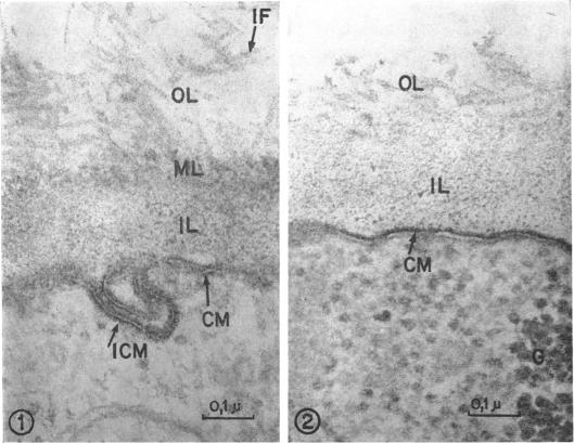
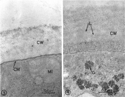
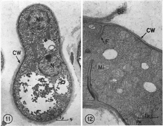
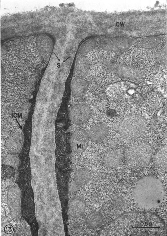
The difference between the budding process of Paracoccidioides brasiliensis and Blastomyces dermatitidis is reported herein. A characteristic feature in P. brasiliensis is that the optical density of the cell wall increases at the site where budding begins and at the neck of the dividing cell, whereas B. dermatitidis does not undergo this alteration. The neck which is formed between the mother and daughter cell at the site of division is much wider in B. dermatitidis than in P. brasiliensis. The bud scar in P. brasiliensis appears as a truncated cone, the top of which is covered only by the inner layer of the cell wall; in comparison, in B. dermatitidis the bud scar exhibits a flattened surface covered by the cell wall. Both fungi show an increase in the number of mitochondria and infoldings of the cytoplasmic membrane at the site of separation, which indicates that at this site there is an increase of metabolic activity.
本文报道了巴西副球孢子菌和皮炎芽生菌出芽过程的差异。巴西副球孢子菌的一个特征是,细胞壁的光密度在出芽开始的部位以及分裂细胞的颈部增加,而皮炎芽生菌则不会发生这种变化。皮炎芽生菌在分裂部位母细胞和子细胞之间形成的颈部比巴西副球孢子菌宽得多。巴西副球孢子菌的芽痕呈截顶圆锥状,其顶部仅被细胞壁的内层覆盖;相比之下,皮炎芽生菌的芽痕呈现出被细胞壁覆盖的扁平表面。两种真菌在分离部位的线粒体数量和细胞质膜内褶都增加,这表明在该部位代谢活动增强。