• 文献检索
  • 文档翻译
  • 深度研究
  • 学术资讯
  • Suppr Zotero 插件Zotero 插件
  • 邀请有礼
  • 套餐&价格
  • 历史记录
应用&插件
Suppr Zotero 插件Zotero 插件浏览器插件Mac 客户端Windows 客户端微信小程序
定价
高级版会员购买积分包购买API积分包
服务
文献检索文档翻译深度研究API 文档MCP 服务
关于我们
关于 Suppr公司介绍联系我们用户协议隐私条款
关注我们

Suppr 超能文献

核心技术专利:CN118964589B侵权必究
粤ICP备2023148730 号-1Suppr @ 2026

文献检索

告别复杂PubMed语法,用中文像聊天一样搜索,搜遍4000万医学文献。AI智能推荐,让科研检索更轻松。

立即免费搜索

文件翻译

保留排版,准确专业,支持PDF/Word/PPT等文件格式,支持 12+语言互译。

免费翻译文档

深度研究

AI帮你快速写综述,25分钟生成高质量综述,智能提取关键信息,辅助科研写作。

立即免费体验

巴西副球孢子菌和皮炎芽生菌的菌丝体阶段:一项电子显微镜研究。

Mycelial phase of Paracoccidioides brasiliensis and Blastomyces dermatitidis: an electron microscope study.

作者信息

Carbonell L M, Rodriguez J

出版信息

J Bacteriol. 1968 Aug;96(2):533-43. doi: 10.1128/jb.96.2.533-543.1968.

DOI:10.1128/jb.96.2.533-543.1968
PMID:5674059
原文链接:https://pmc.ncbi.nlm.nih.gov/articles/PMC252327/
Abstract

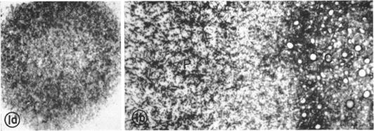
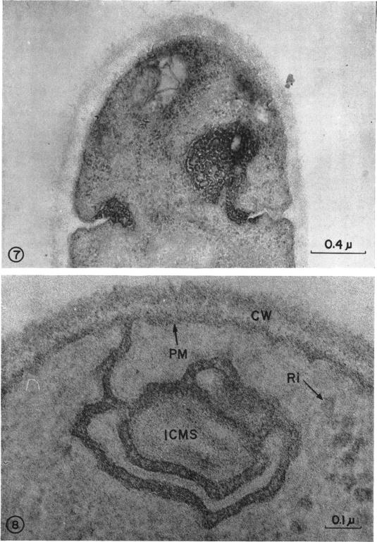
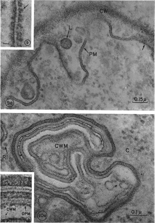
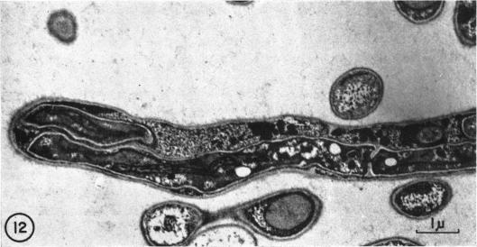
A comparative study of the mycelial phase of Paracoccidioides brasiliensis and Blastomyces dermatitidis reveals that both fungi are very much alike, containing multiple nuclei and nuclear pores, mitochondria, ribosomes, scarce endoplasmic reticulum, intracytoplasmic membrane systems, glycogen, and vacuoles. Shadowed cell walls show fine fibrillar surfaces that contrast with those in the yeast phase. The intracytoplasmic membrane system is continuous with the plasma membrane and is similar to bacterial mesosomes. Granules with light cores and dark rims are observed in the plasma membrane. Live hyphae growing inside a dead hypha are found much more frequently in immersed cultures than in solid-medium cultures, suggesting that breakage of the hypha could elicit this phenomenon.

摘要

巴西副球孢子菌和皮炎芽生菌菌丝体阶段的比较研究表明,这两种真菌非常相似,都含有多个细胞核和核孔、线粒体、核糖体、稀少的内质网、胞质内膜系统、糖原和液泡。经投射处理的细胞壁显示出精细的纤维状表面,这与酵母阶段的细胞壁表面形成对比。胞质内膜系统与质膜相连,类似于细菌间体。在质膜中观察到有浅色核心和深色边缘的颗粒。在浸没培养物中比在固体培养基培养物中更频繁地发现活菌丝在死菌丝内生长,这表明菌丝的断裂可能引发这种现象。

https://cdn.ncbi.nlm.nih.gov/pmc/blobs/9f42/252327/b31f81da201c/jbacter00395-0295-a.jpg
https://cdn.ncbi.nlm.nih.gov/pmc/blobs/9f42/252327/5fdd952a2731/jbacter00395-0288-a.jpg
https://cdn.ncbi.nlm.nih.gov/pmc/blobs/9f42/252327/786f21e11739/jbacter00395-0289-a.jpg
https://cdn.ncbi.nlm.nih.gov/pmc/blobs/9f42/252327/183d4abf97ab/jbacter00395-0290-a.jpg
https://cdn.ncbi.nlm.nih.gov/pmc/blobs/9f42/252327/1bc74b7ecdfd/jbacter00395-0291-a.jpg
https://cdn.ncbi.nlm.nih.gov/pmc/blobs/9f42/252327/02a2fc218ef3/jbacter00395-0293-a.jpg
https://cdn.ncbi.nlm.nih.gov/pmc/blobs/9f42/252327/36e2365a3878/jbacter00395-0294-a.jpg
https://cdn.ncbi.nlm.nih.gov/pmc/blobs/9f42/252327/5c23bc7ca6fa/jbacter00395-0294-b.jpg
https://cdn.ncbi.nlm.nih.gov/pmc/blobs/9f42/252327/b31f81da201c/jbacter00395-0295-a.jpg

相似文献

1
Mycelial phase of Paracoccidioides brasiliensis and Blastomyces dermatitidis: an electron microscope study.巴西副球孢子菌和皮炎芽生菌的菌丝体阶段:一项电子显微镜研究。
J Bacteriol. 1968 Aug;96(2):533-43. doi: 10.1128/jb.96.2.533-543.1968.
2
Cell wall changes during the budding process of Paracoccidioides brasiliensis and Blastomyces dermatitidis.巴西副球孢子菌和皮炎芽生菌出芽过程中的细胞壁变化。
J Bacteriol. 1967 Jul;94(1):213-23. doi: 10.1128/jb.94.1.213-223.1967.
3
Intrayeast hyphae in Paracoccidioides brasiliensis.巴西副球孢子菌中的酵母内菌丝。
J Bacteriol. 1969 Jun;98(3):1395-6. doi: 10.1128/jb.98.3.1395-1396.1969.
4
Mycelial- to yeast-phase transitions of the dimorphic fungi Blastomyces dermatitidis and Paracoccidioides brasiliensis.双相真菌皮炎芽生菌和巴西副球孢子菌的菌丝体到酵母相转变。
J Bacteriol. 1987 Sep;169(9):4055-60. doi: 10.1128/jb.169.9.4055-4060.1987.
5
The fine structure of the microconidium of Blastomyces dermatitidis.
Mycopathol Mycol Appl. 1974 Feb 28;52(2):93-100. doi: 10.1007/BF02128051.
6
Ultrastructural changes during the yeastlike to mycelial-phase conversion of Blastomyces dermatitidis and Histoplasma capsulatum.皮炎芽生菌和荚膜组织胞浆菌从酵母样向菌丝相转化过程中的超微结构变化。
J Bacteriol. 1970 Feb;101(2):628-35. doi: 10.1128/jb.101.2.628-635.1970.
7
Electron microscopy of centrifuged hyphae of Neurospora.粗糙脉孢菌离心菌丝的电子显微镜观察。
J Biophys Biochem Cytol. 1961 Mar;9(3):609-17. doi: 10.1083/jcb.9.3.609.
8
Electronmicroscopy of self-parasitism by Histoplasma capsulatum and Blastomyces dermatitidis.荚膜组织胞浆菌和皮炎芽生菌自身寄生现象的电子显微镜观察
Mycopathol Mycol Appl. 1970 Aug 31;40(3):271-6. doi: 10.1007/BF02051780.
9
Ultrastructural studies on the dimorphism of Paracoccidioides brasiliensis, Blastomyces dermatitidis and Histoplasma capsulatum.巴西副球孢子菌、皮炎芽生菌和荚膜组织胞浆菌二态性的超微结构研究。
Sabouraudia. 1981 Dec;19(4):275-86. doi: 10.1080/00362178185380451.
10
Granuloma formation and killing functions of granuloma in congenitally athymic nude mice infected with Blastomyces dermatitidis and Paracoccidioides brasiliensis.感染皮炎芽生菌和巴西副球孢子菌的先天性无胸腺裸鼠中肉芽肿的形成及肉芽肿的杀伤功能
Mycopathologia. 1983 Jun 20;82(3):129-41. doi: 10.1007/BF00439218.

引用本文的文献

1
The paracoccidioides cell wall: past and present layers toward understanding interaction with the host.副球孢子菌细胞壁:了解与宿主相互作用的过去与现在层面
Front Microbiol. 2011 Dec 20;2:257. doi: 10.3389/fmicb.2011.00257. eCollection 2011.
2
The PbMDJ1 gene belongs to a conserved MDJ1/LON locus in thermodimorphic pathogenic fungi and encodes a heat shock protein that localizes to both the mitochondria and cell wall of Paracoccidioides brasiliensis.PbMDJ1基因属于嗜热双态致病真菌中保守的MDJ1/LON基因座,编码一种热休克蛋白,该蛋白定位于巴西副球孢子菌的线粒体和细胞壁。
Eukaryot Cell. 2006 Feb;5(2):379-90. doi: 10.1128/EC.5.2.379-390.2006.
3

本文引用的文献

1
Electron microscopy of Neurospora crassa mycelia.粗糙脉孢菌菌丝体的电子显微镜观察。
J Biophys Biochem Cytol. 1959 Dec;6(3):423-6. doi: 10.1083/jcb.6.3.423.
2
A stain for fungi in tissue sections and smears using Gomori's methenamine-silver nitrate technic.一种使用戈莫里甲萘胺银硝酸盐技术对组织切片和涂片进行真菌染色的方法。
Am J Clin Pathol. 1955 Aug;25(8):975-9. doi: 10.1093/ajcp/25.8_ts.0975.
3
TRANSFORMATION OF MYCELIAL AND YEAST FORMS OF PARACOCCIDIOIDES BRASILIENSIS IN CULTURES AND IN EXPERIMENTAL INOCULATIONS.巴西副球孢子菌在培养物中和实验接种中的菌丝体及酵母形态转化
Blastomycoma of the cerebellum. An ultrastructural study.
小脑母细胞瘤。一项超微结构研究。
Acta Neuropathol. 1980;50(2):109-14. doi: 10.1007/BF00692860.
4
The cell wall of fungal human pathogens: its possible role in host-parasite relationships.人类致病真菌的细胞壁:其在宿主-寄生虫关系中的可能作用。
Mycopathologia. 1982 Sep 17;79(3):159-84. doi: 10.1007/BF01837196.
5
Estrogens inhibit mycelium-to-yeast transformation in the fungus Paracoccidioides brasiliensis: implications for resistance of females to paracoccidioidomycosis.雌激素抑制巴西副球孢子菌中菌丝体向酵母的转化:对女性抵抗副球孢子菌病的意义。
Infect Immun. 1984 Nov;46(2):346-53. doi: 10.1128/iai.46.2.346-353.1984.
6
Intrayeast hyphae in Paracoccidioides brasiliensis.巴西副球孢子菌中的酵母内菌丝。
J Bacteriol. 1969 Jun;98(3):1395-6. doi: 10.1128/jb.98.3.1395-1396.1969.
7
Cell wall composition of the yeast and mycelial forms of Paracoccidioides brasiliensis.巴西副球孢子菌酵母型和菌丝体型的细胞壁组成
J Bacteriol. 1969 Mar;97(3):1036-41. doi: 10.1128/jb.97.3.1036-1041.1969.
8
Electronmicroscopy of self-parasitism by Histoplasma capsulatum and Blastomyces dermatitidis.荚膜组织胞浆菌和皮炎芽生菌自身寄生现象的电子显微镜观察
Mycopathol Mycol Appl. 1970 Aug 31;40(3):271-6. doi: 10.1007/BF02051780.
9
Ultrastructural changes during the yeastlike to mycelial-phase conversion of Blastomyces dermatitidis and Histoplasma capsulatum.皮炎芽生菌和荚膜组织胞浆菌从酵母样向菌丝相转化过程中的超微结构变化。
J Bacteriol. 1970 Feb;101(2):628-35. doi: 10.1128/jb.101.2.628-635.1970.
10
Ultrastructure of dimorphic transformation in paracoccidioides brasiliensis.巴西副球孢子菌双态转变的超微结构
J Bacteriol. 1969 Nov;100(2):1076-82. doi: 10.1128/jb.100.2.1076-1082.1969.
J Bacteriol. 1965 Aug;90(2):504-10. doi: 10.1128/jb.90.2.504-510.1965.
4
FINE STRUCTURE OF FUNGI AS REVEALED BY ELECTRON MICROSCOPY.电子显微镜揭示的真菌精细结构
Biol Rev Camb Philos Soc. 1965 Feb;40:52-92. doi: 10.1111/j.1469-185x.1965.tb00795.x.
5
[ULTRASTRUCTURE OF PARACOCCIDIOIDES BRASILIENSIS IN CULTURES OF THE YEAST-LIKE PHASE].[巴西副球孢子菌酵母样相培养物的超微结构]
Mycopathol Mycol Appl. 1963 Jun 15;19:184-204. doi: 10.1007/BF02051247.
6
Formation of intracytoplasmic membrane system of mycobacteria related to cell division.分枝杆菌胞质内膜系统的形成与细胞分裂相关。
J Bacteriol. 1963 Jan;85(1):150-63. doi: 10.1128/jb.85.1.150-163.1963.
7
A new epoxy embedment for electron microscopy.一种用于电子显微镜的新型环氧树脂包埋剂。
J Cell Biol. 1962 Jun;13(3):437-43. doi: 10.1083/jcb.13.3.437.
8
"Myelin figures" in yeast cultures of Paracoccidioides brasiliensis.巴西副球孢子菌酵母培养物中的“髓鞘样结构”
J Bacteriol. 1962 Jun;83(6):1356-7. doi: 10.1128/jb.83.6.1356-1357.1962.
9
Relation between basophilia and fine structure of cytoplasm in the fungus Allomyces macrogynus Em.大雌异水霉(Allomyces macrogynus Em.)中嗜碱性与细胞质精细结构的关系
J Biophys Biochem Cytol. 1960 Feb;7(1):127-34. doi: 10.1083/jcb.7.1.127.
10
[Continuity of plasma membrane and nuclear membrane].[质膜与核膜的连续性]
Exp Cell Res. 1959 Mar;16(3):689-92. doi: 10.1016/0014-4827(59)90139-9.