Scheidtmann K H, Echle B, Walter G
J Virol. 1982 Oct;44(1):116-33. doi: 10.1128/JVI.44.1.116-133.1982.
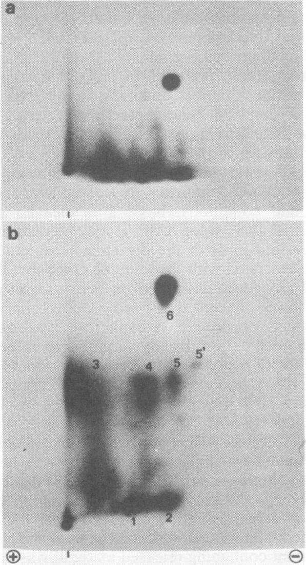
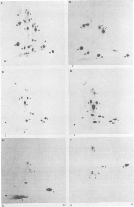
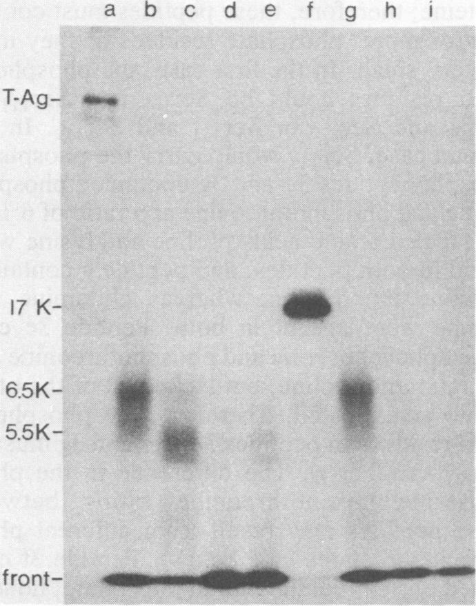
The phosphorylation sites of simian virus 40 large T antigen were determined within the primary structure of the molecule. Exhaustive digestion of (32)P-labeled large T antigen with trypsin generated six major phosphopeptides which could be separated in a newly developed isobutyric acid-containing chromatography system. By partial tryptic digestion, large T antigen was cleaved into an amino-terminal fragment of 17,000 daltons and overlapping fragments from the carboxy-terminal region ranging in size between 71,000 and 13,000 daltons. The location of the phosphopeptides was then determined by fingerprint analyses of individual fragments. Their physical properties were analyzed by sizing on polyacrylamide gels and by sequential digestion and peptide mapping; their amino acid composition was determined by differential labeling with various amino acids. The amino-terminal 17,000-dalton fragment gave rise to only one phosphopeptide (phosphopeptide 3) that contained half of the phosphate label incorporated into large T antigen. It contained phosphoserine and phosphothreonine sites, all of which were clustered within a small segment between Cys(105) and Lys(127). This segment contained five serines and two threonines. Among these, Ser(106), Ser(123), and Thr(124) were identified as phosphorylated residues; in addition, either one or both of Ser(111) and Ser(112) were phosphorylated. The neighboring residues, Ser(123) and Thr(124), were found in three different phosphorylation states in that either Ser(123) or Thr(124) or both were phosphorylated. Phosphopeptides 1, 2, 4, 5, and 6 were all derived from a single fragment extending 26,000 daltons upstream from the carboxy terminus of large T antigen. Phosphopeptide 6 was identical with the previously determined phosphothreonine peptide phosphorylated at Thr(701). Phosphopeptides 1, 2, 4, and 5 contained only serine-bound phosphate. Phosphopeptides 1, 2, and 4 represented overlapping peptides, all of which were phosphorylated at Ser(639) located next to a cluster of six acidic residues. In phosphopeptide 5, a large peptide ranging from Asn(653) to Arg(691), at least two of seven serines were phosphorylated. Thus, large T antigen contains at least eight phosphorylation sites. Their clustering within two separate regions might correlate with structural and functional domains of this protein.
在猴病毒40大T抗原分子的一级结构中确定了其磷酸化位点。用胰蛋白酶对(32)P标记的大T抗原进行彻底消化,产生了六个主要的磷酸肽,这些磷酸肽可以在新开发的含异丁酸的色谱系统中分离。通过部分胰蛋白酶消化,大T抗原被切割成一个17,000道尔顿的氨基末端片段和来自羧基末端区域的重叠片段,大小在71,000至13,000道尔顿之间。然后通过对各个片段的指纹分析来确定磷酸肽的位置。通过在聚丙烯酰胺凝胶上进行大小分析以及顺序消化和肽图谱分析来分析它们的物理性质;通过用各种氨基酸进行差异标记来确定它们的氨基酸组成。氨基末端的17,000道尔顿片段只产生了一个磷酸肽(磷酸肽3),该磷酸肽包含了掺入大T抗原中的一半磷酸标记。它含有磷酸丝氨酸和磷酸苏氨酸位点,所有这些位点都聚集在Cys(105)和Lys(127)之间的一个小片段内。这个片段包含五个丝氨酸和两个苏氨酸。其中,Ser(106)、Ser(123)和Thr(124)被鉴定为磷酸化残基;此外,Ser(111)和Ser(112)中的一个或两个被磷酸化。相邻的残基Ser(123)和Thr(124)处于三种不同磷酸化状态,即要么Ser(123)要么Thr(124)或者两者都被磷酸化。磷酸肽1、2、4、5和6都来自一个从大T抗原羧基末端向上游延伸26,000道尔顿的单一片段。磷酸肽6与先前确定的在Thr(701)处磷酸化的磷酸苏氨酸肽相同。磷酸肽1、2、4和5只含有与丝氨酸结合的磷酸。磷酸肽1、2和4代表重叠肽,它们都在紧邻六个酸性残基簇的Ser(639)处被磷酸化。在磷酸肽5中,一个从Asn(653)到Arg(691)的大肽段中,七个丝氨酸中的至少两个被磷酸化。因此,大T抗原至少含有八个磷酸化位点。它们在两个不同区域内的聚集可能与该蛋白质的结构和功能域相关。